Electroencephalography (EEG) is a non-invasive neuroimaging technique that measures the electrical activity of the brain through electrodes placed on the scalp. EEG can be used for various research applications, including studying brain function and activity, identifying neurological disorders, and investigating the effects of drugs or other interventions on brain activity. EEG is particularly useful for studying brain activity in real-time and identifying the timing and location of brain activity associated with specific cognitive processes or behaviors. It can also be used in clinical settings to diagnose and monitor neurological disorders such as epilepsy, sleep disorders, and traumatic brain injuries. Additionally, EEG can be used to investigate the effects of various interventions, such as cognitive training or neurofeedback, on brain activity and function.
For surfers, catching the perfect wave can induce a state of pure ecstasy known as the “stoke”. But what’s happening in the brain during this ultimate ride? Wearable Sensing created a custom dry EEG system that measures brainwaves during surfing. They partnered with Red Bull to use this technology on professional surfers to uncover the neurophysiological aspects of surfing. The dry EEG system is worn on the head like a swimming cap, and it allows for the measurement of brain activity in real-time during surfing. By studying the brainwaves of surfers during their best rides, researchers hope to understand what goes on in the brain during moments of flow and peak performance, and ultimately unlock the secrets to achieving that elusive state of “stoke”.
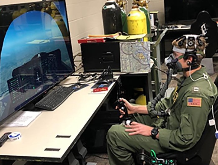
In this study, wearable sensors and machine learning-based algorithms were used to predict hypoxia in-flight. The group used Wearable Sensing’s dry-EEG technology to collect sensor data from 85 participants during a two-phase study. Participants wore aviation flight masks, which regulated their oxygen intake while performing cognitive tests and simulated flying tasks. EEG data was collected and analyzed using principal component analysis and machine learning algorithms, including Naïve Bayes, decision tree, random forest, and neural network algorithms, to classify the data as normal or hypoxic. The results showed high sensitivity and specificity, indicating potential for developing a real-time, in-flight hypoxia detection system.
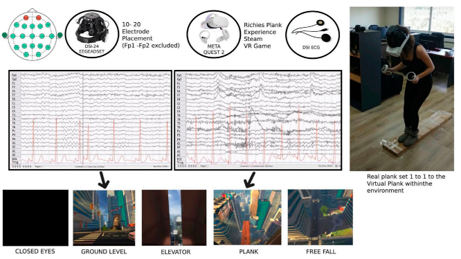
This paper proposes a protocol for assessing stress using wearable sensing technology, including Electroencephalography (EEG), Electrocardiography (ECG), and the Perceived Stress Scale, in combination with a Virtual Reality phobia induction setting. Wearable Sensing’s dry EEG technology is used to measure brain activity and investigate functional brain connectivity associated with stress. The proposed protocol can be expanded with the incorporation of machine learning algorithms for automatic stress level classification.
Rahimi-Nasrabadi, Hamed; Jin, Jianzhong; Mazade, Reece; Pons, Carmen; Najafian, Sohrab; Alonso, Jose-Manuel
Image luminance changes contrast sensitivity in visual cortex Journal Article
In: Cell reports, vol. 34, no. 5, pp. 108692, 2021.
@article{rahimi2021image,

title = {Image luminance changes contrast sensitivity in visual cortex},

author = {Hamed Rahimi-Nasrabadi and Jianzhong Jin and Reece Mazade and Carmen Pons and Sohrab Najafian and Jose-Manuel Alonso},

doi = {https://doi.org/10.1016/j.celrep.2021.108692},

year  = {2021},

date = {2021-02-02},

journal = {Cell reports},

volume = {34},

number = {5},

pages = {108692},

publisher = {Elsevier},

abstract = {Accurate measures of contrast sensitivity are important for evaluating visual disease progression and for navigation safety. Previous measures suggested that cortical contrast sensitivity was constant across widely different luminance ranges experienced indoors and outdoors. Against this notion, here, we show that luminance range changes contrast sensitivity in both cat and human cortex, and the changes are different for dark and light stimuli. As luminance range increases, contrast sensitivity increases more within cortical pathways signaling lights than those signaling darks. Conversely, when the luminance range is constant, light-dark differences in contrast sensitivity remain relatively constant even if background luminance changes. We show that a Naka-Rushton function modified to include luminance range and light-dark polarity accurately replicates both the statistics of light-dark features in natural scenes and the cortical responses to multiple combinations of contrast and luminance. We conclude that differences in light-dark contrast increase with luminance range and are largest in bright environments.},

keywords = {},

pubstate = {published},

tppubtype = {article}

}
Neilson, Brittany N; Phillips, Jeffrey B; Snider, Dallas H; Drollinger, Sabrina M; Linnville, Steven E; Mayes, Ryan S
A Data-Driven Approach to Aid in Understanding Brainwave Activity During Hypoxia Conference
2020 IEEE Research and Applications of Photonics in Defense Conference (RAPID), IEEE IEEE, Miramar Beach, FL, USA, 2020, ISBN: 978-1-7281-5890-7.
@conference{neilson2020data,

title = {A Data-Driven Approach to Aid in Understanding Brainwave Activity During Hypoxia},

author = {Brittany N Neilson and Jeffrey B Phillips and Dallas H Snider and Sabrina M Drollinger and Steven E Linnville and Ryan S Mayes},

doi = {10.1109/RAPID49481.2020.9195700},

isbn = {978-1-7281-5890-7},

year  = {2020},

date = {2020-09-14},

booktitle = {2020 IEEE Research and Applications of Photonics in Defense Conference (RAPID)},

pages = {1--2},

publisher = {IEEE},

address = {Miramar Beach, FL, USA},

organization = {IEEE},

abstract = {Changes in brainwave activity have been associated with hypoxia, but the literature is inconsistent. Twenty-five participants were subjected to normobaric hypoxia while undergoing a variety of cognitive tasks. The detected differences in brain activity between normal and hypoxic conditions are presented.},

keywords = {},

pubstate = {published},

tppubtype = {conference}

}
Haar Millo, S; Faisal, A
Brain Activity Reveals Multiple Motor-Learning Mechanisms in a Real-World Task Journal Article
In: Frontiers in Human Neuroscience, vol. 14, pp. 354, 2020, ISBN: 1662-5161.
@article{haarbrain,

title = {Brain Activity Reveals Multiple Motor-Learning Mechanisms in a Real-World Task},

author = {Haar Millo, S and Faisal, A},

doi = {10.3389/fnhum.2020.00354},

isbn = {1662-5161},

year  = {2020},

date = {2020-09-02},

journal = {Frontiers in Human Neuroscience},

volume = {14},

pages = {354},

publisher = {Frontiers Media},

abstract = {Many recent studies found signatures of motor learning in neural beta oscillations (13–30 Hz), and specifically in the post-movement beta rebound (PMBR). All these studies were in controlled laboratory-tasks in which the task designed to induce the studied learning mechanism. Interestingly, these studies reported opposing dynamics of the PMBR magnitude over learning for the error-based and reward-based tasks (increase vs. decrease, respectively). Here, we explored the PMBR dynamics during real-world motor-skill-learning in a billiards task using mobile-brain-imaging. Our EEG recordings highlight the opposing dynamics of PMBR magnitudes (increase vs. decrease) between different subjects performing the same task. The groups of subjects, defined by their neural dynamics, also showed behavioral differences expected for different learning mechanisms. Our results suggest that when faced with the complexity of the real-world different subjects might use different learning mechanisms for the same complex task. We speculate that all subjects combine multi-modal mechanisms of learning, but different subjects have different predominant learning mechanisms.},

keywords = {},

pubstate = {published},

tppubtype = {article}

}
Kim, Young-June; Park, Jin-Hong; Cho, Young-Suk; Kim, Keum-Sook
In: Journal of Convergence for Information Technology, vol. 10, no. 8, pp. 203–212, 2020.
@article{kim2020effect,

title = {The Effect of Cognitive Rehabilitation Program Using Virtual Reality (VR) Contents on Cognitive function, Depression, Upper Extremity Function and Activities of Daily Living in the Elderly},

author = {Young-June Kim and Jin-Hong Park and Young-Suk Cho and Keum-Sook Kim},

url = {https://www.koreascience.or.kr/article/JAKO202024852036461.page},

year  = {2020},

date = {2020-08-28},

journal = {Journal of Convergence for Information Technology},

volume = {10},

number = {8},

pages = {203--212},

publisher = {Convergence Society for SMB},

abstract = {The purpose of this study was to investigate the effects of cognitive rehabilitation programs using Virtual Reality(VR) content on the daily living abilities such as cognitive abilities, depression, and upper extremity functions of the elderly. The study group analyzed the effectiveness by separating the experimental group, which is the virtual reality cognitive rehabilitation application group, and the control group, the universal cognitive stimulation program application group. As a result of the study, the MMSE-K score improved by 13.0% in the experimental group and 2.3% in the control group. The improvement in each area of the experimental group was found to be 3.1% MBI, 7.1% MFT(Rt.), 3.5% MFT(Lt.), and 25.4% K-GDS. As a result of comparing the pre-post score change between each group, there was a significant difference between groups in daily living ability (p<.001) and MFT(Rt.)(p<.01). In addition, as a result of comparing the changes in absolute alpha waves to confirm the degree of depression through brain waves, there was no statistically significant difference. However, in the experimental group, it was confirmed that the average value increased to a positive value. This study is an experiment to verify the effectiveness of the cognitive rehabilitation program using virtual reality contents, and suggests a new intervention method to maintain and improve the daily life ability, cognitive function, depression and upper extremity function of the elderly.},

keywords = {},

pubstate = {published},

tppubtype = {article}

}
Lim, Hyunmi; Kim, Won-Seok; Ku, Jeonghun
Transcranial Direct Current Stimulation Effect on Virtual Hand Illusion Journal Article
In: Cyberpsychology, Behavior, and Social Networking, vol. 23, no. 8, pp. 541–549, 2020.
@article{lim2020transcranial,

title = {Transcranial Direct Current Stimulation Effect on Virtual Hand Illusion},

author = {Hyunmi Lim and Won-Seok Kim and Jeonghun Ku},

doi = {https://doi.org/10.1089/cyber.2019.0741},

year  = {2020},

date = {2020-08-04},

urldate = {2020-08-04},

journal = {Cyberpsychology, Behavior, and Social Networking},

volume = {23},

number = {8},

pages = {541--549},

publisher = {Mary Ann Liebert, Inc., publishers 140 Huguenot Street, 3rd Floor New~…},

abstract = {Virtual reality (VR) is effectively used to evoke the mirror illusion, and transcranial direct current stimulation (tDCS) synergistically facilitates this illusion. This study investigated whether a mirror virtual hand illusion (MVHI) induced by an immersive, first-person-perspective, virtual mirror system could be modulated by tDCS of the primary motor cortex. Fourteen healthy adults (average age 21.86 years ±0.47, seven men and seven women) participated in this study, and they experienced VR with and without tDCS—the tDCS and sham conditions, each of which takes ∼30 minutes—on separate days to allow the washout of the tDCS effect. While experiencing VR, the movements of the virtual left hand reflected the flexion and extension of the real right hand. Subsequently, electroencephalogram was recorded, the magnitude of the proprioceptive shift was measured, and the participants provided responses to a questionnaire regarding hand ownership. A significant difference in the proprioceptive shift was observed between the tDCS and sham conditions. In addition, there was significant suppression of the mu power in Pz, and augmentation of the beta power in the Pz, P4, O1, and O2 channels. The difference in proprioceptive deviation between the two conditions showed significant negative correlation with mu suppression over the left frontal lobe in the tDCS condition. Finally, the question “I felt that the virtual hand was my own hand” received a significantly higher score under the tDCS condition. In short, applying tDCS over the motor cortex facilitates the MVHI by activating the attentional network over the parietal and frontal lobes such that the MVHI induces more proprioceptive drift, which suggests that the combination of VR and tDCS can enhance the immersive effect in VR. This result provides better support for the use of the MVHI paradigm in combination with tDCS for recovery from illnesses such as stroke.},

keywords = {},

pubstate = {published},

tppubtype = {article}

}
Son, Ji Eun; Choi, Hyoseon; Lim, Hyunmi; Ku, Jeonghun
In: Technology and Health Care, vol. 28, no. S1, pp. 509-519, 2020.
@article{son2020development,

title = {Development of a flickering action video based steady state visual evoked potential triggered brain computer interface-functional electrical stimulation for a rehabilitative action observation game},

author = {Ji Eun Son and Hyoseon Choi and Hyunmi Lim and Jeonghun Ku},

editor = {Severin P. Schwarzacher and Carlos Gómez},

doi = {10.3233/THC-209051},

year  = {2020},

date = {2020-06-04},

journal = {Technology and Health Care},

volume = {28},

number = {S1},

pages = {509-519},

publisher = {IOS Press},

abstract = {BACKGROUND:

This study focused on developing an upper limb rehabilitation program. In this regard, a steady state visual evoked potential (SSVEP) triggered brain computer interface (BCI)-functional electrical stimulation (FES) based action observation game featuring a flickering action video was designed.


OBJECTIVE:

In particular, the synergetic effect of the game was investigated by combining the action observation paradigm with BCI based FES.


METHODS:

The BCI-FES system was contrasted under two conditions: with flickering action video and flickering noise video. In this regard, 11 right-handed subjects aged between 22–27 years were recruited. The differences in brain activation in response to the two conditions were examined.


RESULTS:

The results indicate that T3 and P3 channels exhibited greater Mu suppression in 8–13 Hz for the action video than the noise video. Furthermore, T4, C4, and P4 channels indicated augmented high beta (21–30 Hz) for the action in contrast to the noise video. Finally, T4 indicated suppressed low beta (14–20 Hz) for the action video in contrast to the noise video.


CONCLUSION:

The flickering action video based BCI-FES system induced a more synergetic effect on cortical activation than the flickering noise based system.},

keywords = {},

pubstate = {published},

tppubtype = {article}

}
Wang, Jiahui; Antonenko, Pavlo; Keil, Andreas; Dawson, Kara
Converging subjective and psychophysiological measures of cognitive load to study the effects of instructor-present video Journal Article
In: Mind, Brain, and Education, vol. 14, no. 3, pp. 279–291, 2020.
@article{wang2020converging,

title = {Converging subjective and psychophysiological measures of cognitive load to study the effects of instructor-present video},

author = {Jiahui Wang and Pavlo Antonenko and Andreas Keil and Kara Dawson},

doi = {https://doi.org/10.1111/mbe.12239},

year  = {2020},

date = {2020-03-30},

urldate = {2020-01-01},

journal = {Mind, Brain, and Education},

volume = {14},

number = {3},

pages = {279--291},

publisher = {Wiley Online Library},

abstract = {Many online videos feature an instructor on the screen to improve learners' engagement; however, the influence of this design on learners' cognitive load is underexplored. This study investigates the effects of instructor presence on learners' processing of information using both subjective and psychophysiological measures of cognitive load. Sixty university students watched a statistics instructional video either with or without instructor presence, while the spontaneous electrical activity of their brain was recorded using electroencephalography (EEG). At the conclusion of the video, they also self-reported overall load, intrinsic load, extraneous load, and germane load they experienced during the video. Learning from the video was assessed via tests of retention and transfer. Results suggested the instructor-present video improved learners' ability to transfer information and was associated with a lower self-reported intrinsic and extraneous load. Event-related changes in theta band activity also indicated lower cognitive load with instructor-present video.},

keywords = {},

pubstate = {published},

tppubtype = {article}

}
Mahdid, Yacine; Lee, Uncheol; Blain-Moraes, Stefanie
Assessing the Quality of Wearable EEG Systems Using Functional Connectivity Journal Article
In: IEEE Access, vol. 8, pp. 193214–193225, 2020, ISSN: 2169-3536.
@article{mahdid2020assessing,

title = {Assessing the Quality of Wearable EEG Systems Using Functional Connectivity},

author = {Yacine Mahdid and Uncheol Lee and Stefanie Blain-Moraes},

doi = {10.1109/ACCESS.2020.3033472},

issn = {2169-3536},

year  = {2020},

date = {2020-01-01},

journal = {IEEE Access},

volume = {8},

pages = {193214--193225},

publisher = {IEEE},

abstract = {Assessing the data quality of wearable electroencephalogram (EEG) systems is critical to collecting reliable neurophysiological data in non-laboratory environments. To date, measures of signal quality and spectral characteristics have been used to characterize wearable EEG systems. We demonstrate that these traditional measures do not provide fine-grained differentiation between the performance of four popular wearable EEG systems (the Epoc+, OpenBCI, DSI-24 and Quick-30 Dry EEG). Using two computationally inexpensive metrics of undirected functional connectivity (phase lag index) and directed functional connectivity (directed phase lag index), we compare the integrity of the phase relationships captured by wearable systems to those recorded from a high-density research-grade EEG system (Electrical Geodesics Inc). Our results demonstrate that functional connectivity analyses provide additional discriminatory information about wearable EEG systems, with clear differentiation of the cosine similarity between research-grade functional connectivity patterns and those generated by each wearable system. We provide a freely available Matlab toolbox containing all metrics described in this paper such that researchers and non-experts interested in wearable EEG systems can easily assess the quality of systems not characterized in this study, thus advancing the translation of EEG research into non-laboratory settings.},

keywords = {},

pubstate = {published},

tppubtype = {article}

}
Islam, Md Shafiqul; El-Hajj, Ahmad M; Alawieh, Hussein; Dawy, Zaher; Abbas, Nabil; El-Imad, Jamil
EEG mobility artifact removal for ambulatory epileptic seizure prediction applications Journal Article
In: Biomedical Signal Processing and Control, vol. 55, pp. 101638, 2020, ISSN: 1746-8094.
@article{islam2020eeg,

title = {EEG mobility artifact removal for ambulatory epileptic seizure prediction applications},

author = {Md Shafiqul Islam and Ahmad M El-Hajj and Hussein Alawieh and Zaher Dawy and Nabil Abbas and Jamil El-Imad},

doi = {https://doi.org/10.1016/j.bspc.2019.101638},

issn = {1746-8094},

year  = {2020},

date = {2020-01-01},

journal = {Biomedical Signal Processing and Control},

volume = {55},

pages = {101638},

publisher = {Elsevier},

abstract = {Mobile monitoring of electroencephalogram (EEG) signals is prone to different sources of artifacts. Most importantly, motion-related artifacts present a major challenge hindering the clean acquisition of EEG data as they spread all over the scalp and across all frequency bands. This leads to additional complexity in the development of neurologically-oriented mobile health solutions. Among the top five most common neurological disorders, epilepsy has increasingly relied on EEG for diagnosis. Separate methods have been used to classify EEG segments in the context of epilepsy while reducing the existing mobility artifacts. This work specifically devises an approach to remove motion-related artifacts in the context of epilepsy. The proposed approach first includes the recording of EEG signals using a wearable EEG headset. The recorded signals are then colored by some motion artifacts generated in a lab-controlled experiment. This stage is followed by temporal and spectral characterization of the signals and artifact removal using independent component analysis (ICA). The proposed approach is tested using real clinical EEG data and results showed an average increase in accuracy of ∼9% in seizure detection and ∼24% in prediction.},

keywords = {},

pubstate = {published},

tppubtype = {article}

}
Choi, Hyoseon; Lim, Hyunmi; Kim, Joon Woo; Kang, Youn Joo; Ku, Jeonghun
Brain computer interface-based action observation game enhances mu suppression in patients with stroke Journal Article
In: Electronics, vol. 8, no. 12, pp. 1466, 2019.
@article{choi2019brain,

title = {Brain computer interface-based action observation game enhances mu suppression in patients with stroke},

author = {Hyoseon Choi and Hyunmi Lim and Joon Woo Kim and Youn Joo Kang and Jeonghun Ku},

doi = {https://doi.org/10.3390/electronics8121466},

year  = {2019},

date = {2019-12-02},

journal = {Electronics},

volume = {8},

number = {12},

pages = {1466},

publisher = {Multidisciplinary Digital Publishing Institute},

abstract = {Action observation (AO), based on the mirror neuron theory, is a promising strategy to promote motor cortical activation in neurorehabilitation. Brain computer interface (BCI) can detect a user’s intention and provide them with brain state-dependent feedback to assist with patient rehabilitation. We investigated the effects of a combined BCI-AO game on power of mu band attenuation in stroke patients. Nineteen patients with subacute stroke were recruited. A BCI-AO game provided real-time feedback to participants regarding their attention to a flickering action video using steady-state visual-evoked potentials. All participants watched a video of repetitive grasping actions under two conditions: (1) BCI-AO game and (2) conventional AO, in random order. In the BCI-AO game, feedback on participants’ observation scores and observation time was provided. In conventional AO, a non-flickering video and no feedback were provided. The magnitude of mu suppression in the central motor, temporal, parietal, and occipital areas was significantly higher in the BCI-AO game than in the conventional AO. The magnitude of mu suppression was significantly higher in the BCI-AO game than in the conventional AO both in the affected and unaffected hemispheres. These results support the facilitatory effects of the BCI-AO game on mu suppression over conventional AO},

keywords = {},

pubstate = {published},

tppubtype = {article}

}
Please fill out the form and provide a brief description of your application so we can help match you with products that will meet your specific needs.
Please fill out the form and provide a brief description of your application so we can help match you with products that will meet your specific needs.
Please fill out the form and provide a brief description of your application so we can help match you with products that will meet your specific needs.