Electroencephalography (EEG) is a non-invasive neuroimaging technique that measures the electrical activity of the brain through electrodes placed on the scalp. EEG can be used for various research applications, including studying brain function and activity, identifying neurological disorders, and investigating the effects of drugs or other interventions on brain activity. EEG is particularly useful for studying brain activity in real-time and identifying the timing and location of brain activity associated with specific cognitive processes or behaviors. It can also be used in clinical settings to diagnose and monitor neurological disorders such as epilepsy, sleep disorders, and traumatic brain injuries. Additionally, EEG can be used to investigate the effects of various interventions, such as cognitive training or neurofeedback, on brain activity and function.
For surfers, catching the perfect wave can induce a state of pure ecstasy known as the “stoke”. But what’s happening in the brain during this ultimate ride? Wearable Sensing created a custom dry EEG system that measures brainwaves during surfing. They partnered with Red Bull to use this technology on professional surfers to uncover the neurophysiological aspects of surfing. The dry EEG system is worn on the head like a swimming cap, and it allows for the measurement of brain activity in real-time during surfing. By studying the brainwaves of surfers during their best rides, researchers hope to understand what goes on in the brain during moments of flow and peak performance, and ultimately unlock the secrets to achieving that elusive state of “stoke”.
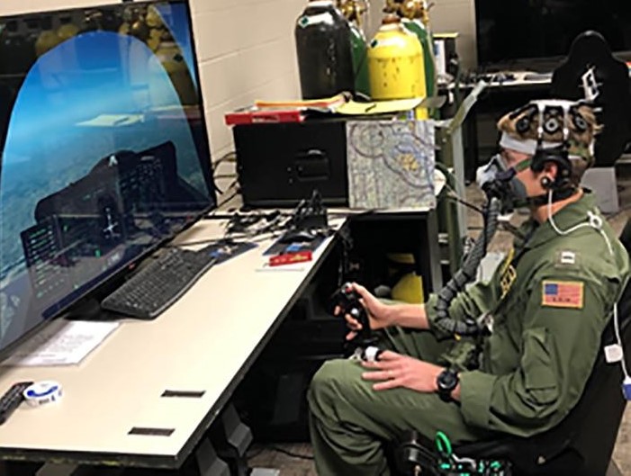
In this study, wearable sensors and machine learning-based algorithms were used to predict hypoxia in-flight. The group used Wearable Sensing’s dry-EEG technology to collect sensor data from 85 participants during a two-phase study. Participants wore aviation flight masks, which regulated their oxygen intake while performing cognitive tests and simulated flying tasks. EEG data was collected and analyzed using principal component analysis and machine learning algorithms, including Naïve Bayes, decision tree, random forest, and neural network algorithms, to classify the data as normal or hypoxic. The results showed high sensitivity and specificity, indicating potential for developing a real-time, in-flight hypoxia detection system.
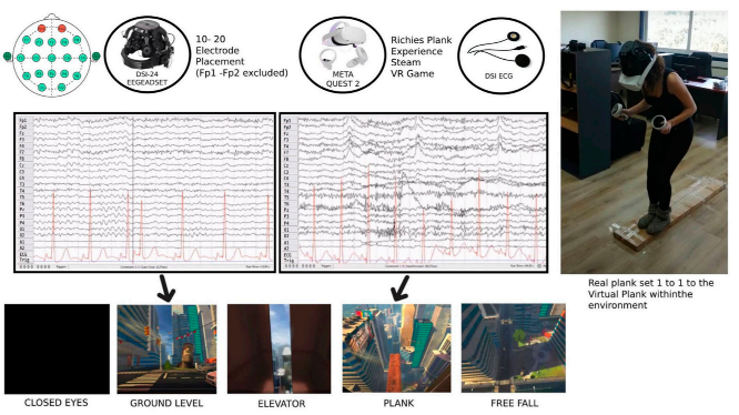
This paper proposes a protocol for assessing stress using wearable sensing technology, including Electroencephalography (EEG), Electrocardiography (ECG), and the Perceived Stress Scale, in combination with a Virtual Reality phobia induction setting. Wearable Sensing’s dry EEG technology is used to measure brain activity and investigate functional brain connectivity associated with stress. The proposed protocol can be expanded with the incorporation of machine learning algorithms for automatic stress level classification.
Raj, Anil; Roberts, Brooke; Hollingshead, Kristy; McDonald, Neil; Poquette, Melissa; Soussou, Walid
A Wearable Multisensory, Multiagent Approach for Detection and Mitigation of Acute Cognitive Strain Conference
International Conference on Augmented Cognition, Springer 2018.
@conference{raj2018wearable,

title = {A Wearable Multisensory, Multiagent Approach for Detection and Mitigation of Acute Cognitive Strain},

author = {Anil Raj and Brooke Roberts and Kristy Hollingshead and Neil McDonald and Melissa Poquette and Walid Soussou},

url = {https://link.springer.com/chapter/10.1007/978-3-319-91470-1_16},

year  = {2018},

date = {2018-06-03},

booktitle = {International Conference on Augmented Cognition},

pages = {180--200},

organization = {Springer},

abstract = {While operators performing tasks with high workload can increase task performance in response to limited increases in cognitive stress, chronic or rapidly accelerating stress can exceed the operator’s ability to compensate, generating acute cognitive strain (ACS). ACS represents a state wherein performance, situation awareness and cooperativity deteriorate markedly, leading to critical errors, mishaps or casualties. Nearly two decades of augmented cognition (AugCog) research has demonstrated the utility of psychophysiologic sensing and analysis for identification and tracking of changes in cognitive state and to modulate human machine interactions for improving system task performance. The proposed approach leveraged prior efforts to modulate cognitive stress using a multiagent approach to acquire and analyze multiple Psychophysiologic sensory channels, including changes in vocalizations, to create a reliable and non-intrusive Detector of Acute Cognitive Strain (DACS). The DACS system provides an integrated wearable multi-modal Research Sensor Suite (RSS) using the open-source Adaptive Multiagent Integration (AMI) architecture, that includes analysis agents for electroencephalograph (EEG), electromyography (EMG), video oculography (VOG), vocalization, and others to identify and correlate physiological signatures with cognitive stress and strain. An online AMI agent-based processing algorithm was developed and applied to audio communications to evaluate for changes in speaker vocalization fundamental frequency (F0) and cadence (utterances per minute). This paper describes initial phase results of aerospace mishap vocalization stress marker detection, a potential element of the proposed DACS system. DACS could use these markers to trigger adaptive automation agents that reduce task load and allow pilots to prevent or recover from ACS episodes.},

keywords = {},

pubstate = {published},

tppubtype = {conference}

}
Vergeer, Mark; Mesik, Juraj; Baek, Yihwa; Wilmerding, Kelton; Engel, Stephen A
Orientation-selective contrast adaptation measured with SSVEP Journal Article
In: Journal of Vision, vol. 18, no. 5, pp. 2–2, 2018.
@article{vergeer2018orientation,

title = {Orientation-selective contrast adaptation measured with SSVEP},

author = {Mark Vergeer and Juraj Mesik and Yihwa Baek and Kelton Wilmerding and Stephen A Engel},

doi = {https://doi.org/10.1167/18.5.2},

year  = {2018},

date = {2018-05-01},

journal = {Journal of Vision},

volume = {18},

number = {5},

pages = {2--2},

publisher = {The Association for Research in Vision and Ophthalmology},

abstract = {Exposure to oriented luminance contrast patterns causes a reduction in visual sensitivity specifically for the adapter orientation. This orientation selectivity is probably the most studied aspect of contrast adaptation, but it has rarely been measured with steady-state visually evoked potentials (SSVEPs), despite their becoming one of the more popular methods of human neuroscience. Here, we measured orientation selective adaptation by presenting a plaid stimulus of which the horizontal and vertical grating reversed contrast at different temporal frequencies, while recording EEG signals from occipital visual areas. In three experiments, we compared SSVEP responses to the plaid before and after adaptation. All experiments showed a significant decrease in SSVEP response at the frequency of the adapter orientation, whereas such an effect was absent for the frequency of the orthogonal orientation. Adaptation also led to robust phase delays, selectively for the SSVEP frequency corresponding to the adapter orientation. These results demonstrate the efficiency of SSVEPs for measuring orientation selective adaptation; the method can measure changes in both amplitude and phase, simultaneously for two orientations.},

keywords = {},

pubstate = {published},

tppubtype = {article}

}
Memmott, Tab; Eddy, Brandon; Dabiri, Sina; Erdogmus, Deniz; Fried-Oken, Melanie; Oken, Barry
Automated and self-report measures of drowsiness over successive calibrations in a brain-computer interface for communication Journal Article
In: Clinical Neurophysiology, vol. 129, pp. e61–e62, 2018.
@article{memmott2018t154,

title = {Automated and self-report measures of drowsiness over successive calibrations in a brain-computer interface for communication},

author = {Tab Memmott and Brandon Eddy and Sina Dabiri and Deniz Erdogmus and Melanie Fried-Oken and Barry Oken},

doi = {https://doi.org/10.1016/j.clinph.2018.04.155},

year  = {2018},

date = {2018-05-01},

journal = {Clinical Neurophysiology},

volume = {129},

pages = {e61--e62},

publisher = {Elsevier},

abstract = {Brain computer interfaces (BCI) generally require the user to maintain an attentive state. Potential end-users with severe speech and physical impairments may have limited communication abilities to report their current state, thus an automatic calculation of state may improve performance. It’s not yet known if an effective automatic calculation of drowsiness can be detected reliably in end-user populations or healthy controls.},

keywords = {},

pubstate = {published},

tppubtype = {article}

}
Arakaki, Xianghong; Shoga, Michael; Li, Lianyang; Zouridakis, George; Tran, Thao; Fonteh, Alfred N; Dawlaty, Jessica; Goldweber, Robert; Pogoda, Janice M; Harrington, Michael G
Alpha desynchronization/synchronization during working memory testing is compromised in acute mild traumatic brain injury (mTBI) Journal Article
In: PloS one, vol. 13, no. 2, pp. e0188101, 2018.
@article{arakaki2018alpha,

title = {Alpha desynchronization/synchronization during working memory testing is compromised in acute mild traumatic brain injury (mTBI)},

author = {Xianghong Arakaki and Michael Shoga and Lianyang Li and George Zouridakis and Thao Tran and Alfred N Fonteh and Jessica Dawlaty and Robert Goldweber and Janice M Pogoda and Michael G Harrington},

doi = {https://doi.org/10.1371/journal.pone.0188101},

year  = {2018},

date = {2018-02-14},

journal = {PloS one},

volume = {13},

number = {2},

pages = {e0188101},

publisher = {Public Library of Science San Francisco, CA USA},

abstract = {Diagnosing and monitoring recovery of patients with mild traumatic brain injury (mTBI) is challenging because of the lack of objective, quantitative measures. Diagnosis is based on description of injuries often not witnessed, subtle neurocognitive symptoms, and neuropsychological testing. Since working memory (WM) is at the center of cognitive functions impaired in mTBI, this study was designed to define objective quantitative electroencephalographic (qEEG) measures of WM processing that may correlate with cognitive changes associated with acute mTBI. First-time mTBI patients and mild peripheral (limb) trauma controls without head injury were recruited from the emergency department. WM was assessed by a continuous performance task (N-back). EEG recordings were obtained during N-back testing on three occasions: within five days, two weeks, and one month after injury. Compared with controls, mTBI patients showed abnormal induced and evoked alpha activity including event-related desynchronization (ERD) and synchronization (ERS). For induced alpha power, TBI patients had excessive frontal ERD on their first and third visit. For evoked alpha, mTBI patients had lower parietal ERD/ERS at the second and third visits. These exploratory qEEG findings offer new and non-invasive candidate measures to characterize the evolution of injury over the first month, with potential to provide much-needed objective measures of brain dysfunction to diagnose and monitor the consequences of mTBI.},

keywords = {},

pubstate = {published},

tppubtype = {article}

}
Hunter, Aimee M; Nghiem, Thien X; Cook, Ian A; Krantz, David E; Minzenberg, Michael J; Leuchter, Andrew F
In: Clinical EEG and Neuroscience, vol. 49, no. 5, pp. 306–315, 2017.
@article{hunter2018change,

title = {Change in quantitative EEG theta cordance as a potential predictor of repetitive transcranial magnetic stimulation clinical outcome in major depressive disorder},

author = {Aimee M Hunter and Thien X Nghiem and Ian A Cook and David E Krantz and Michael J Minzenberg and Andrew F Leuchter},

doi = {https://doi.org/10.1177/1550059417746212},

year  = {2017},

date = {2017-12-10},

urldate = {2017-12-10},

journal = {Clinical EEG and Neuroscience},

volume = {49},

number = {5},

pages = {306--315},

publisher = {Sage Publications Sage CA: Los Angeles, CA},

abstract = {Repetitive transcranial magnetic stimulation (rTMS) has demonstrated efficacy in major depressive disorder (MDD), although clinical outcome is variable. Change in the resting-state quantitative electroencephalogram (qEEG), particularly in theta cordance early in the course of treatment, has been linked to antidepressant medication outcomes but has not been examined extensively in clinical rTMS. This study examined change in theta cordance over the first week of clinical rTMS and sought to identify a biomarker that would predict outcome at the end of 6 weeks of treatment. Clinically stable outpatients (n = 18) received nonblinded rTMS treatment administered to the dorsolateral prefrontal cortex (DLPFC). Treatment parameters (site, intensity, number of pulses) were adjusted on an ongoing basis guided by changes in symptom severity rating scale scores. qEEGs were recorded at pretreatment baseline and after 1 week of left DLPFC (L-DLPFC) rTMS using a 21-channel dry-electrode headset. Analyses examined the association between week 1 regional changes in theta band (4-8 Hz) cordance, and week 6 patient- and physician-rated outcomes. Theta cordance change in the central brain region predicted percent change in Inventory of Depressive Symptomology–Self-Report (IDS-SR) score, and improvement versus nonimprovement on the Clinical Global Impression–Improvement Inventory (CGI-I) (R2 = .38, P = .007; and Nagelkerke R2 = .78, P = .0001, respectively). The cordance biomarker remained significant when controlling for age, gender, and baseline severity. Treatment-emergent change in EEG theta cordance in the first week of rTMS may predict acute (6-week) treatment outcome in MDD. This oscillatory synchrony biomarker merits further study in independent samples.},

keywords = {},

pubstate = {published},

tppubtype = {article}

}
Mills, Caitlin; Fridman, Igor; Soussou, Walid; Waghray, Disha; Olney, Andrew M; D'Mello, Sidney K
Put your thinking cap on: detecting cognitive load using EEG during learning Conference
Proceedings of the Seventh International Learning Analytics & Knowledge Conference, 2017.
@conference{mills2017put,

title = {Put your thinking cap on: detecting cognitive load using EEG during learning},

author = {Caitlin Mills and Igor Fridman and Walid Soussou and Disha Waghray and Andrew M Olney and Sidney K D'Mello},

doi = {https://doi.org/10.1145/3027385.3027431},

year  = {2017},

date = {2017-03-01},

booktitle = {Proceedings of the Seventh International Learning Analytics & Knowledge Conference},

pages = {80--89},

abstract = {Current learning technologies have no direct way to assess students' mental effort: are they in deep thought, struggling to overcome an impasse, or are they zoned out? To address this challenge, we propose the use of EEG-based cognitive load detectors during learning. Despite its potential, EEG has not yet been utilized as a way to optimize instructional strategies. We take an initial step towards this goal by assessing how experimentally manipulated (easy and difficult) sections of an intelligent tutoring system (ITS) influenced EEG-based estimates of students' cognitive load. We found a main effect of task difficulty on EEG-based cognitive load estimates, which were also correlated with learning performance. Our results show that EEG can be a viable source of data to model learners' mental states across a 90-minute session.},

keywords = {},

pubstate = {published},

tppubtype = {conference}

}
Halford, Jonathan J; Schalkoff, Robert J; Satterfield, Kevin E; Martz, Gabriel U; Kutluay, Ekrem; Waters, Chad G; Dean, Brian C
Comparison of a Novel Dry Electrode Headset to Standard Routine EEG in Veterans Journal Article
In: Journal of Clinical Neurophysiology, vol. 33, no. 6, pp. 530–537, 2016.
@article{halford2016comparison,

title = {Comparison of a Novel Dry Electrode Headset to Standard Routine EEG in Veterans},

author = {Jonathan J Halford and Robert J Schalkoff and Kevin E Satterfield and Gabriel U Martz and Ekrem Kutluay and Chad G Waters and Brian C Dean},

doi = {10.1097/WNP.0000000000000284},

year  = {2016},

date = {2016-12-01},

journal = {Journal of Clinical Neurophysiology},

volume = {33},

number = {6},

pages = {530--537},

publisher = {LWW},

abstract = {Objective:

This purpose of this study was to evaluate the usefulness of a prototype battery-powered dry electrode system (DES) EEG recording headset in Veteran patients by comparing it with standard EEG.


Methods:

Twenty-one Veterans had both a standard electrode system recording and DES recording in nine different patient states at the same encounter. Setup time, patient comfort, and subject preference were measured. Three experts performed technical quality rating of each EEG recording in a blinded fashion using the web-based EEGnet system. Power spectra were compared between DES and standard electrode system recordings.


Results:

The average time for DES setup was 5.7 minutes versus 21.1 minutes for standard electrode system. Subjects reported that the DES was more comfortable during setup. Most subjects (15 of 21) preferred the DES. On a five-point scale (1—best quality to 5—worst quality), the technical quality of the standard electrode system recordings was significantly better than for the DES recordings, at 1.25 versus 2.41 (P < 0.0001). But experts found that 87% of the DES EEG segments were of sufficient technical quality to be interpretable.


Conclusions:

This DES offers quick and easy setup and is well tolerated by subjects. Although the technical quality of DES recordings was less than standard EEG, most of the DES recordings were rated as interpretable by experts.


Significance:

This DES, if improved, could be useful for a telemedicine approach to outpatient routine EEG recording within the Veterans Administration or other health system.},

keywords = {},

pubstate = {published},

tppubtype = {article}

}
Fridman, Igor; Cordeiro, Malaika; Rais-Bahrami, Khodayar; McDonald, Neil J; Jr, James J Reese; Massaro, An N; Conry, Joan A; Chang, Taeun; Soussou, Walid; Tsuchida, Tammy N
Evaluation of dry sensors for neonatal EEG recordings Journal Article
In: Journal of clinical neurophysiology: official publication of the American Electroencephalographic Society, vol. 33, no. 2, pp. 149, 2016.
@article{fridman2016evaluation,

title = {Evaluation of dry sensors for neonatal EEG recordings},

author = {Igor Fridman and Malaika Cordeiro and Khodayar Rais-Bahrami and Neil J McDonald and James J Reese Jr and An N Massaro and Joan A Conry and Taeun Chang and Walid Soussou and Tammy N Tsuchida},

url = {https://www.ncbi.nlm.nih.gov/pmc/articles/PMC4818163/},

year  = {2016},

date = {2016-04-01},

journal = {Journal of clinical neurophysiology: official publication of the American Electroencephalographic Society},

volume = {33},

number = {2},

pages = {149},

publisher = {NIH Public Access},

abstract = {Introduction

Neonatal seizures are a common neurologic diagnosis in Neonatal Intensive Care Units (NICUs), occurring in approximately 14,000 newborns annually in the US. While the only reliable means of detecting and treating neonatal seizures is with an EEG recording, many neonates do not get an EEG or experience delays in getting them. Barriers to obtaining neonatal EEGs include: 1) lack of skilled EEG technologists to apply conventional wet electrodes to delicate neonatal skin, 2) poor signal quality due to improper skin preparation and artifact, 3) extensive time needed to apply electrodes. Dry sensors have the potential to overcome these obstacles but have not been previously evaluated on neonates.


Methods

Sequential and simultaneous recordings with wet and dry sensors were performed for one hour on 27 neonates from 35-42.5 weeks postmenstrual age. Recordings were analyzed for correlation and amplitude, and were reviewed by neurophysiologists. Performance of dry sensors on simulated vernix was examined.


Results

Analysis of dry and wet signals showed good time-domain correlation (reaching >0.8) given the non-superimposed sensor positions, and similar power spectral density curves. Neurophysiologist reviews showed no statistically significant difference between dry and wet data on most clinically-relevant EEG background and seizure patterns. There was no skin injury after 1 hr of dry sensor recordings. In contrast to wet electrodes, impedance and electrical artifact of dry sensors were largely unaffected by simulated vernix.


Conclusions

Dry sensors evaluated in this study have the potential to provide high-quality, timely EEG recordings on neonates with less risk of skin injury.},

keywords = {},

pubstate = {published},

tppubtype = {article}

}
Arakaki, Xianghong; Shoga, Michael; Li, Lianyang; Zouridakis, George; Rostami, Ramona; Goldweber, Robert; Harrington, Michael
Exploring neuroplasticity in acute mild traumatic brain injury Journal Article
In: The FASEB Journal, vol. 30, pp. 992–4, 2016.
@article{arakaki2016exploring,

title = {Exploring neuroplasticity in acute mild traumatic brain injury},

author = {Xianghong Arakaki and Michael Shoga and Lianyang Li and George Zouridakis and Ramona Rostami and Robert Goldweber and Michael Harrington},

url = {https://faseb.onlinelibrary.wiley.com/doi/abs/10.1096/fasebj.30.1_supplement.992.4},

year  = {2016},

date = {2016-04-01},

journal = {The FASEB Journal},

volume = {30},

pages = {992--4},

publisher = {The Federation of American Societies for Experimental Biology},

abstract = {Objectives

To explore neuroplasticity in a longitudinal study of acute mild traumatic brain injury (mTBI).


Methods

We are using quantitative electroencephalography (qEEG) and magnetoencephalography (MEG) during the resting state and during cognitive brain stress to explore neuroplasticity in an ongoing acute mild traumatic brain injury research. Acute mTBI patients are recruited from the emergency department of Huntington Memorial Hospital in Pasadena, CA, and controls are non‐head‐trauma patients. Brain stress includes the N‐back (0‐back and 2‐back) working memory test and Color‐Word Interference Test (CWIT), administered using E‐prime software. Data were collected at three time points: within 1 week of injury, 14 days, and 30 days after injury. Behavioral as well as MEG and qEEG analysis are performed to compare the two groups.


Results

Resting MEG detected low frequency activity in the mTBI group, consistent with previous publications. N‐back, in particular during 2‐back trials, and CWIT, in particular during incongruent trials, both show initial executive function impairment that improved on later visits. Time frequency analysis suggested corresponding compromised brain activity.


Conclusions

The EEG/MEG recordings during rest and brain stress are objective and sensitive to neuroplasticity in acute mTBI, and could be potential objective mTBI markers.},

keywords = {},

pubstate = {published},

tppubtype = {article}

}
Kang, Dayoon; Kim, Jinsoo; Jang, Dong-Pyo; Cho, Yang Seok; Kim, Sung-Phil
Investigation of engagement of viewers in movie trailers using electroencephalography Journal Article
In: Brain-Computer Interfaces, vol. 2, no. 4, pp. 193–201, 2015.
@article{kang2015investigation,

title = {Investigation of engagement of viewers in movie trailers using electroencephalography},

author = {Dayoon Kang and Jinsoo Kim and Dong-Pyo Jang and Yang Seok Cho and Sung-Phil Kim},

doi = {https://doi.org/10.1080/2326263X.2015.1103591},

year  = {2015},

date = {2015-11-10},

journal = {Brain-Computer Interfaces},

volume = {2},

number = {4},

pages = {193--201},

publisher = {Taylor & Francis},

abstract = {Brain-computer interfaces (BCIs) have been focused on providing direct communications to the disabled. Recently, BCI researchers have expanded BCI applications to non-medical uses and categorized them as active BCI, reactive BCI, and passive BCI. Neurocinematics, a new application of reactive BCIs, aims to understand viewers’ cognitive and affective responses to movies from neural activity, providing more objective information than traditional subjective self-reports. However, studies on analytical indices for neurocinematics have verified their indices by comparisons with self-reports. To overcome this contradictory issue, we proposed using an independent psychophysical index to evaluate a neural engagement index (NEI). We made use of the secondary task reaction time (STRT), which measures participants’ engagement in a primary task by their reaction time to a secondary task; here, responding to a tactile stimulus was the secondary task and watching a movie trailer was the primary task. NEI was developed as changes in the difference between frontal beta and alpha activity of EEG. We evaluated movie trailers using NEI, STRT, and self-reports and found a significant correlation between STRT and NEI across trailers but no correlation between any of the self-report results and STRT or NEI. Our results suggest that NEI developed for neurocinematics may conform well with more objective psychophysical assessments but not with subjective self-reports.},

keywords = {},

pubstate = {published},

tppubtype = {article}

}
Please fill out the form and provide a brief description of your application so we can help match you with products that will meet your specific needs.
Please fill out the form and provide a brief description of your application so we can help match you with products that will meet your specific needs.
Please fill out the form and provide a brief description of your application so we can help match you with products that will meet your specific needs.