Wearable Sensing’s wireless DSI-24 is the leading dry electrode EEG system in terms of signal quality and comfort. The DSI-24 takes on average less than 3 minutes to set up, making it the ideal solution for scientists in need of a simple, easy to use, EEG system. Our patented sensor technology not only delivers uncompromised signal quality but also enables our system to be virtually immune against motion and electrical artifacts. As a result, the DSI-24 can be utilized in virtual or augmented reality, while also allowing researchers to take their experiments out of the lab, and into the real world.
The DSI-24 has sensors that provide full head coverage with 19 electrodes on the head, 2 earclip sensors, and also has 3 built-in auxiliary inputs for acquisition of up to 3 auxiliary sensors. It also has an 8-bit trigger input to synchronize with other devices such as Eye-Tracking, Motion (IMU), and more.
Used around the world by leaders in Research, Neurofeedback, Neuromarketing, Brain-Computer Interfaces, & Neuroergonomics.
With over 90% correlation to research-grade wet EEG systems, the dry sensor interface (DSI) offers unparalleled quality and performance
Multiple adjustment points and a foam pad lined interior enable the system to be worn for up to 8 hours on any head shape or size
All DSI systems include free, unlimited licenses of DSI-Streamer, our data acquisition software which can record raw data, in .csv and .edf file formats
Faraday cage's, spring-loaded electrodes, and our patented common-mode follower technology, provides near immunity against electrical and motion artifacts
Using 70% isopropyl alcohol and a cleaning brush, the DSI-24 only takes a minute to clean, 3 minutes to dry, and can be up and running on the next subject in minutes
All DSI systems include our free C based .dll API, which enables users to pull the raw data directly from the headset, for custom software on Windows, Mac OS, Linux, and ARM
The DSI-24 was designed for ultra-rapid setup, taking on average less than 3 minutes to don, and works on any type of hair, including long hair, thick hair, afros, and more
DSI headsets have active sensors, amplifiers, digitizers, batteries, onboard storage, and wireless transmission, making them complete, mobile, wearable EEG systems
DSI systems exclusively work with QStates, a machine learning algorithm for cognitive classification on states such as mental workload, engagement, and fatigue
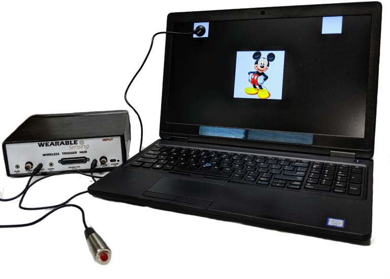
Our Wireless Trigger Hub simplifies the synchronization of DSI headsets with other devices. It features:
An additional benefit of the Trigger Hub design is that it allows synchronization across multiple data sources that are distributed across multiple systems, each of which running at its own clock rate. One such case commonly experienced in EEG experiments involves the synchronization of EEG and eye-tracking measurements, where the inevitable clock drift that arises between two systems during extended measurements creates difficulty in aligning data to events across the two systems.
The DSI-24 has 3 auxiliary inputs on the headset, which allows for automatic synchronization of Wearable Sensing’s auxiliary sensors to the EEG. The sensors available include ECG, EMG, EOG, GSR, RESP, & TEMP. The sensor data is collected and recorded in our data acquisition software, DSI-Streamer, where you can view the EEG and Aux sensors in real-time.
EEG Channels
Fp1, Fp2, Fz, F3, F4, F7, F8, Cz, C3, C4, T7/T3, T8/T4, Pz, P3, P4, P7/T5, P8/T6, O1, O2, A1, A2
Reference / Ground
Common Mode Follower / Fpz
Head Size Range
Adult Size: 52cm – 62cm circumference
Child Size: 48cm – 54cm circumference
Sampling Rate
300 Hz (600Hz upgrade available)
Bandwidth
0.003 – 150 Hz
A/D resolution
0.317 μV referred to input
Input Impedance (1Hz)
47 GΩ
CMRR
> 120 dB
Amplifier / Digitizer
16 bits / 24 channels
Wireless
Bluetooth
Wireless Range
10 m
Run-time
> 24 Hours, Hot-Swappable Batteries
Onboard Storage
~ 68 Hours (available option)
Data Acquisition
Real time, evoked potentials
Signal Quality Monitoring
Continuous impedance, Baseline offset, Noise (1-50 Hz)
Data Type
Raw and Filtered Data available
File Type
.CSV and .EDF
Data Output Streaming
TCP/IP socket, API (C Based), LSL
Cognitive State Classification
Brain Computer Interface
SSVEP BCI Algorithms; BCI2000; OpenViBE; PsychoPy; BCILab
Data Integration / Analysis
CAPTIV; Lab Streaming Layer; NeuroPype; BrainStorm; NeuroVIS
Neurofeedback
Applied Neuroscience NeuroGuide; Brainmaster Brain Avatar; EEGer
Neuromarketing
CAPTIV Neurolab
Presentation
Presentation; E-Prime
Kambhamettu, Sudhendra; Cruz, Meenalosini Vimal; Anitha, S; Chakkaravarthy, S Sibi; Kumar, K Nandeesh
Brain-Computer Interface-Assisted Automated Wheelchair Control Management--Cerebro: A BCI Application Journal Article
In: Brain-Computer Interface: Using Deep Learning Applications, pp. 205–229, 2023.
@article{kambhamettu2023brain,

title = {Brain-Computer Interface-Assisted Automated Wheelchair Control Management--Cerebro: A BCI Application},

author = {Sudhendra Kambhamettu and Meenalosini Vimal Cruz and S Anitha and S Sibi Chakkaravarthy and K Nandeesh Kumar},

doi = {https://doi.org/10.1002/9781119857655.ch9},

year  = {2023},

date = {2023-02-10},

urldate = {2023-01-01},

journal = {Brain-Computer Interface: Using Deep Learning Applications},

pages = {205--229},

publisher = {Wiley Online Library},

abstract = {Technology today serves millions of people suffering from mobility impairments across the globe in numerous ways. Although advancements in medicine and healthcare systems improve the life expectancy of the general population, sophisticated engineering techniques and computing processes have long facilitated the patient in the recovery process. People struggling with mobility impairments and especially spine injuries which also leads to loss of speech, often have a narrow group of devices to aid them move from place-to-place and they are often limited to just movement functionality. BCI (Brain Computer Interface) powered wheelchairs leverage the power of the brain, i.e. translating the thoughts/neural activity into real-world movement providing automated motion without any third party intervention. Many BCI powered wheelchairs in the market are cumbersome to operate and provide only singular functionality of movement. To address this problem and improve the state of BCI products, Cerebro introduces the first ever go-to market product utilizing Artificial Intelligence to facilitate mobility features with built-in speech functionality via blink detection. Further sections of the Chapter take an in-depth look into each layer of the Cerebro system.


},

keywords = {},

pubstate = {published},

tppubtype = {article}

}
Dhaliwal, BS; Haddad, J; Debrincat, M; others,
In: Correspondence: Peter Hurwitz, Clarity Science LLC, vol. 750, 2022.
@article{dhaliwal2022changes,

title = {Changes in Electroencephalogram (EEG) After Foot Stimulation with Embedded Haptic Vibrotactile Trigger Technology: Neuromatrix and Pain Modulation Considerations. Anesth Pain Res. 2022; 6 (2): 1-11},

author = {BS Dhaliwal and J Haddad and M Debrincat and others},

url = {https://www.scivisionpub.com/pdfs/improvement-in-balance-and-stability-using-a-novel-sensory-application-haptic-vibrotactile-trigger-technology-2537.pdf},

year  = {2022},

date = {2022-12-16},

urldate = {2022-01-01},

journal = {Correspondence: Peter Hurwitz, Clarity Science LLC},

volume = {750},

abstract = {Background: Globally, pain and pain-related diseases are the leading causes of disability and disease burden. In the United States, pain is the most common reason patients consult primary care providers. An estimated 100 million people live with chronic or recurrent pain. Existing pharmacological treatments for pain include anti-inflammatory agents, opioids, and other oral and topical analgesics. Many of these have been associated with troublesome and potentially harmful adverse effects. Understanding the complex pain neuromatrix may help in identifying alternative, non-invasive strategies and treatment approaches to address pain severity, interference, and improve patient outcomes. The neuromatrix of pain is a network of neuronal pathways and circuits responding to sensory (nociceptive) stimulation. Research has suggested that the output patterns of the body-self neuromatrix are responsible for causing or triggering perceptual, homeostatic, and behavioral programs following traumatic injury, other pathology, or chronic stress. As such, pain can be considered a product of the output of a widely distributed neural network within the brain instead of a sequential result of sensory inputs triggered by injury, inflammation, or other pathology. For over a century, the Brodmann Areas remain the most widely known and frequently cited cytoarchitectural organization of the human cortex. Certain Brodmann areas of the brain have been associated with the current understanding of the neuromatrix of pain. The areas expands well beyond the thalamus and anterior cingulate, and primary (S1) and secondary (S2) somatosensory cortices to include the midbrain region of the periaqueductal gray (PAG) and the lenticular complex as well as the insula, orbitofrontal (Brodmann's area [BA] 11, 47), prefrontal (BA 9, 10, 44-46), motor (BA 6, Supplementary motor area, and M1), inferior parietal (BA 39, 40), and anterior cingulate (BA 24, 25) cortices (ACCs). Treatments that are non-invasive and non-pharmacological and target both central and peripheral nociceptive mechanisms that are identified as having an impact on the Brodmann areas associated with the neuromatrix of pain may potentially be considered a beneficial pain management option for patients. Haptic vibrotactile trigger technology targets the nociceptive pathways and is theorized to disrupt the neuromatrix of pain. The technology has been incorporated into non-pharmacological patches and other non-invasive routes of delivery such as apparel (socks), braces, wristbands, and compression sleeves. The purpose of this minimal risk study was to compare electroencephalogram (EEG) patterns in areas of the brain that have been associated with the neuromatrix for pain in subjects wearing socks that were embedded with haptic vibrotactile trigger technology with those patients that wore socks that were not embedded with the technology.


Methods: This IRB-approved study compared electroencephalogram (EEG) patterns in subjects wearing cloth socks embedded with haptic vibrotactile trigger technology (Superneuro VTT Enhanced Socks (Srysty Holding Co., Toronto, Canada) with those patients that wore cloth socks that were not embedded with the technology. Baseline EEG data from 19 scalp locations were recorded in sixty (60) adult subjects (36 females and 24 males) ranging from ages 14 to 83 wearing standard store-purchased cloth socks on their feet. The subject’s standard socks were then removed and replaced with the Superneuro VTT enhanced socks on the subject’s feet. A second EEG recording was then obtained. Both eyes-closed and eyes-open data were recorded.


Results: The results showed statistically significant t-test differences (P < .01) in 59 out of 60 subjects in absolute power and 60 out of 60 subjects showed statistically significant differences in coherence and phase difference. The largest differences were in the alpha1 and beta2 frequency bands and especially in central scalp locations. Paired t-tests of LORETA current source densities between socks on and socks off demonstrated statistically significant differences in 60 out of 60 subjects. The largest effects of Superneuro VTT enhanced socks on were on the medial bank of the somatosensory cortex as well as in the left frontal lobes in the theta and alpha frequency.


Conclusions: Study results indicate that foot stimulation with embedded haptic vibrotactile trigger technology showed significant modulation in the Brodmann areas that have been shown to be associated with the neuromatrix for pain in the human brain. Further research is suggested to evaluate if this technology has a positive impact on pain severity, pain interference, and quality of life and to be considered as a potentially beneficial pain management strategy and as part of a multi-modal treatment approach. },

keywords = {},

pubstate = {published},

tppubtype = {article}

}
Ocay, Don Daniel; Teel, Elizabeth F; Luo, Owen D; Savignac, Chloé; Mahdid, Yacine; Blain-Moraes, Stefanie; Ferland, Catherine E
Electroencephalographic characteristics of children and adolescents with chronic musculoskeletal pain Journal Article
In: PAIN Reports, vol. 7, no. 6, pp. e1054, 2022.
@article{ocay2022electroencephalographic,

title = {Electroencephalographic characteristics of children and adolescents with chronic musculoskeletal pain},

author = {Don Daniel Ocay and Elizabeth F Teel and Owen D Luo and Chloé Savignac and Yacine Mahdid and Stefanie Blain-Moraes and Catherine E Ferland},

url = {https://journals.lww.com/painrpts/Fulltext/2022/12000/Electroencephalographic_characteristics_of.17.aspx},

year  = {2022},

date = {2022-12-01},

urldate = {2022-12-01},

journal = {PAIN Reports},

volume = {7},

number = {6},

pages = {e1054},

publisher = {LWW},

abstract = {Introduction:

The pathophysiology of pediatric musculoskeletal (MSK) pain is unclear, contributing to persistent challenges to its management.


Objectives:

This study hypothesizes that children and adolescents with chronic MSK pain (CPs) will show differences in electroencephalography (EEG) features at rest and during thermal pain modalities when compared with age-matched controls.


Methods:

One hundred forty-two CP patients and 45 age-matched healthy controls (HCs) underwent a standardized thermal tonic heat and cold stimulations, while a 21-electrode headset collected EEG data. Cohorts were compared with respect to their EEG features of spectral power, peak frequency, permutation entropy, weight phase-lag index, directed phase-lag index, and node degree at 4 frequency bands, namely, delta (1–4 Hz), theta (4–8 Hz), alpha (8–13 Hz), and beta (13–30 Hz), at rest and during the thermal conditions.


Results:

At rest, CPs showed increased global delta (P = 0.0493) and beta (P = 0.0002) power in comparison with HCs. These findings provide further impetus for the investigation and prevention of long-lasting developmental sequalae of early life chronic pain processes. Although no cohort differences in pain intensity scores were found during the thermal pain modalities, CPs and HCs showed significant difference in changes in EEG spectral power, peak frequency, permutation entropy, and network functional connectivity at specific frequency bands (P < 0.05) during the tonic heat and cold stimulations.


Conclusion:

This suggests that EEG can characterize subtle differences in heat and cold pain sensitivity in CPs. The complementation of EEG and evoked pain in the clinical assessment of pediatric chronic MSK pain can better detect underlying pain mechanisms and changes in pain sensitivity.},

keywords = {},

pubstate = {published},

tppubtype = {article}

}
Chang, Won Kee; Lim, Hyunmi; Park, Seo Hyun; Lim, Chaiyoung; Paik, Nam-Jong; Kim, Won-Seok; Ku, Jeonghun
Effect of Immersive Virtual Mirror Visual Feedback on Mu Suppression and Coherence in Motor and Parietal Cortex in Stroke Journal Article
In: 2022.
@article{chang2022effect,

title = {Effect of Immersive Virtual Mirror Visual Feedback on Mu Suppression and Coherence in Motor and Parietal Cortex in Stroke},

author = {Won Kee Chang and Hyunmi Lim and Seo Hyun Park and Chaiyoung Lim and Nam-Jong Paik and Won-Seok Kim and Jeonghun Ku},

doi = {https://doi.org/10.21203/rs.3.rs-2253842/v1},

year  = {2022},

date = {2022-11-11},

urldate = {2022-01-01},

abstract = {Background: This study aimed to investigate the activation pattern of the motor cortex (M1) and parietal cortex during immersive virtual reality (VR)-based mirror visual feedback (MVF) of the upper limb in patients with chronic stroke. Methods: Fourteen patients with chronic stroke with severe upper limb hemiparesis (Brunnstrom stage of hand 1-3) and 21 healthy controls were included. The participants performed wrist extension tasks with their unaffected wrists (or the dominant side in controls). In the MVF condition, the movement of the affected hand was synchronized with that of the unaffected hand. In contrast, only the movement of the unaffected hand was shown in the no-MVF condition. Electroencephalography was obtained during experiments with two conditions (MVF vs no-MVF). Mu suppression in the bilateral M1 and parietal cortex and mu coherence between the ipsilateral M1 and parietal cortex in each hemisphere and interhemispheric M1 were used for analyses. Results: In patients with stroke, MVF induced significant mu suppression in both the ipsilesional M1 and parietal lobes (p=0.006 and p=0.009, respectively), while significant mu suppression was observed in the bilateral M1 (p=0.003 for ipsilesional and p=0.041 for contralesional M1, respectively) and contralesional (contralateral hemisphere to the moving hand) parietal lobes in the healthy controls (p=0.036). The ipsilesional mu coherence between the M1 and parietal cortex in patients with stroke was stronger than that in controls regardless of MVF condition (p<0.001), while mu coherence between interhemispheric M1 cortices was significantly weaker in patients with stroke (p=0.032). Conclusion: In patients with stroke, MVF using immersive VR induces mu suppression in the ipsilesional M1 and parietal lobe. Our findings provide evidence of the neural mechanism of MVF using immersive VR and support its application in patients with stroke with severe hemiparesis.},

keywords = {},

pubstate = {published},

tppubtype = {article}

}
Rustamov, Nabi; Wilson, Elizabeth A; Fogarty, Alexandra E; Crock, Lara W; Leuthardt, Eric C; Haroutounian, Simon
Relief of chronic pain associated with increase in midline frontal theta power Journal Article
In: Pain Reports, vol. 7, no. 6, 2022.
@article{rustamov2022relief,

title = {Relief of chronic pain associated with increase in midline frontal theta power},

author = {Nabi Rustamov and Elizabeth A Wilson and Alexandra E Fogarty and Lara W Crock and Eric C Leuthardt and Simon Haroutounian},

doi = {10.1097/PR9.0000000000001040},

year  = {2022},

date = {2022-10-10},

urldate = {2022-01-01},

journal = {Pain Reports},

volume = {7},

number = {6},

publisher = {Wolters Kluwer Health},

abstract = {Introduction:

There is a need to identify objective cortical electrophysiological correlates for pain relief that could potentially contribute to a better pain management. However, the field of developing brain biomarkers for pain relief is still largely underexplored.


Objectives:

The objective of this study was to investigate cortical electrophysiological correlates associated with relief from chronic pain. Those features of pain relief could serve as potential targets for novel therapeutic interventions to treat pain.


Methods:

In 12 patients with chronic pain in the upper or lower extremity undergoing a clinically indicated nerve block procedure, brain activity was recorded by means of electroencephalogram before and 30 minutes after the nerve block procedure. To determine the specific cortical electrophysiological correlates of relief from chronic pain, 12 healthy participants undergoing cold-pressor test to induce experimental acute pain were used as a control group. The data were analyzed to characterize power spectral density patterns of pain relief and identify their source generators at cortical level.


Results:

Chronic pain relief was associated with significant delta, theta, and alpha power increase at the frontal area. However, only midfrontal theta power increase showed significant positive correlation with magnitude of reduction in pain intensity. The sources of theta power rebound were located in the left dorsolateral prefrontal cortex (DLPFC) and midline frontal cortex. Furthermore, theta power increase in the midline frontal cortex was significantly higher with chronic vs acute pain relief.


Conclusion:

These findings may provide basis for targeting chronic pain relief via modulation of the midline frontal theta oscillations.},

keywords = {},

pubstate = {published},

tppubtype = {article}

}
Woo, Hee-Soon; Song, Chiang-Soon
Effects of Low Visual Acuity Simulations on Eye-Hand Coordination and Brainwaves in Healthy Adults Journal Article
In: Physical Therapy Rehabilitation Science, vol. 11, no. 3, pp. 296–303, 2022.
@article{woo2022effects,

title = {Effects of Low Visual Acuity Simulations on Eye-Hand Coordination and Brainwaves in Healthy Adults},

author = {Hee-Soon Woo and Chiang-Soon Song},

doi = {https://doi.org/10.14474/ptrs.2022.11.3.296},

year  = {2022},

date = {2022-09-30},

urldate = {2022-01-01},

journal = {Physical Therapy Rehabilitation Science},

volume = {11},

number = {3},

pages = {296--303},

publisher = {Korean Academy of Physical Therapy Rehabilitation Science},

abstract = {Objective: In general, macular degeneration, cataracts and glaucoma generally cause visual injury in clinical settings. This study aimed to examine the effects of low visual acuity simulations on hand manual dexterity function and brainwaves in healthy young adults.

Design: Cross-sectional study design

Methods: This study was an observational, cross-sectional study. Seventy healthy young adults participated in this study. To evaluate the effects of low visual acuity simulations on hand function and brain waves, this study involved four different visual conditions including (1) normal vision, (2) simulated cataracts, (3) simulated glaucoma, and (4) simulated macular degeneration. The hand function was measured to use the Minnesota manual dexterity test (MMDT), and the brainwaves was also measured to use the electroencephalography.

Results: In hand function, placing and turning performance on the MMDT in the normal visual condition was significantly different than that in the cataract and macular degeneration conditions (p<0.05), and the placing performance was significantly differred in the normal condition than that in the simulated glaucoma. However, turning was not significantly different in the normal condition than that in the simulated glaucoma. The alpha, beta, and gamma waves did not significantly differ among the four visual conditions (p>0.05).

Conclusions: The results suggest that limited visual information negatively affects the ability to perform tasks requiring arm-hand dexterity and eye-hand coordination. However, the effectiveness of low visual acuity on the brainwaves should be further studied for rehabilitative evidence of visual impairment.},

keywords = {},

pubstate = {published},

tppubtype = {article}

}
Miltiadous, Andreas; Aspiotis, Vasileios; Sakkas, Konstantinos; Giannakeas, Nikolaos; Glavas, Euripidis; Tzallas, Alexandros T
An experimental protocol for exploration of stress in an immersive VR scenario with EEG Conference
2022 7th South-East Europe Design Automation, Computer Engineering, Computer Networks and Social Media Conference (SEEDA-CECNSM), IEEE 2022.
@conference{miltiadous2022experimental,

title = {An experimental protocol for exploration of stress in an immersive VR scenario with EEG},

author = {Andreas Miltiadous and Vasileios Aspiotis and Konstantinos Sakkas and Nikolaos Giannakeas and Euripidis Glavas and Alexandros T Tzallas},

doi = {10.1109/SEEDA-CECNSM57760.2022.9932987},

year  = {2022},

date = {2022-09-23},

urldate = {2022-01-01},

booktitle = {2022 7th South-East Europe Design Automation, Computer Engineering, Computer Networks and Social Media Conference (SEEDA-CECNSM)},

pages = {1--5},

organization = {IEEE},

abstract = {Stress is a subject always relevant to scientific research due to the numerous implications in human life. Typical biomarkers used in the physiological evaluation of stress include Electrocardiography, cortisol levels, galvanic skin response and other. Recently, one less widely used instrument for the assessment of stress that has been re-emerged due to advancements in computational power and machine learning techniques, is Electroencephalography. Moreover, as Virtual Reality HMDs are being rapidly adopted by the research community it becomes apparent that leveraging the offered advantages of VR for the exploration of stress can lead to novel controlable and reproducable experimental procedures. In this paper we combine EEG, ECG and the Perceived Stress Scale with a Virtual Reality phobia induction setting, to propose a protocol for assessing stress. The suggested protocol can be used for functional brain connectivity investigation and thus the evaluation of stress while it and can be expanded via the incorporation of machine learning algorithms for automatic stress level classification.},

keywords = {},

pubstate = {published},

tppubtype = {conference}

}
Rominger, Christian; Gubler, Dani`ele A; Makowski, Lisa M; Troche, Stefan J
In: International journal of psychophysiology, 2022.
@article{rominger2022more,

title = {More creative ideas are associated with increased right posterior power and frontal-parietal/occipital coupling in the upper alpha band: A within-subjects study},

author = {Christian Rominger and Dani`ele A Gubler and Lisa M Makowski and Stefan J Troche},

doi = {https://doi.org/10.1016/j.ijpsycho.2022.08.012},

year  = {2022},

date = {2022-09-01},

urldate = {2022-01-01},

journal = {International journal of psychophysiology},

publisher = {Elsevier},

abstract = {The neurophysiological investigation of creative idea generation is a growing research area. EEG studies congruently reported the sensitivity of upper alpha power (10-12 Hz) for the creative ideation process and its outcome. However, the majority of studies were between-subject design studies and research directly comparing the neurophysiological activation pattern when generating more and less creative ideas within a person are rare. Therefore, the present study was specifically focused on investigating brain activation patterns associated with the generation of more vs. less creative ideas. We applied an alternate uses task (AU-task; i.e., finding original uses for everyday objects such as a brick) in a sample of 74 participants and recorded the brain activation during the AU-task and reference period. A portable EEG system with 21 dry electrodes arranged in the international 10–20 system and linked ear as reference was used. We found a higher increase of upper alpha power during creative ideation (relative to reference period, i.e., task-related power, TRP) over right posterior sites when people generated more compared to less creative ideas. This was accompanied by an increase of functional coupling (i.e., task-related coherence increase) between frontal and parietal/occipital sites, which suggests higher internal attention and more control over sensory processes. Taken together, these findings complement the existing creativity research literature and indicate the importance of alpha power for the creative ideation process also within people.},

keywords = {},

pubstate = {published},

tppubtype = {article}

}
Hu, Yuxia; Wang, Yufei; Zhang, Rui; Hu, Yubo; Fang, Mingzhu; Li, Zhe; Shi, Li; Zhang, Yankun; Zhang, Zhong; Gao, Jinfeng; others,
Assessing stroke rehabilitation degree based on quantitative EEG index and nonlinear parameters Journal Article
In: Cognitive Neurodynamics, pp. 1–9, 2022.
@article{hu2022assessing,

title = {Assessing stroke rehabilitation degree based on quantitative EEG index and nonlinear parameters},

author = {Yuxia Hu and Yufei Wang and Rui Zhang and Yubo Hu and Mingzhu Fang and Zhe Li and Li Shi and Yankun Zhang and Zhong Zhang and Jinfeng Gao and others},

url = {https://link.springer.com/article/10.1007/s11571-022-09849-4},

year  = {2022},

date = {2022-08-06},

urldate = {2022-01-01},

journal = {Cognitive Neurodynamics},

pages = {1--9},

publisher = {Springer},

abstract = {The assessment of motor function is critical to the rehabilitation of stroke patients. However, commonly used evaluation methods are based on behavior scoring, which lacks neurological indicators that directly reflect the motor function of the brain. The objective of this study was to investigate whether resting-state EEG indicators could improve stroke rehabilitation evaluation. We recruited 68 participants and recorded their resting-state EEG data. According to Brunnstrom stage, the participants were divided into three groups: severe, moderate, and mild. Ten quantitative electroencephalographic (QEEG) and five non-linear parameters of resting-state EEG were calculated for further analysis. Statistical tests were performed, and the genetic algorithm-support vector machine was used to select the best feature combination for classification. We found the QEEG parameters show significant differences in Delta, Alpha1, Alpha2, DAR, and DTABR (P < 0.05) among the three groups. Regarding nonlinear parameters, ApEn, SampEn, Lz, and C0 showed significant differences (P < 0.05). The optimal feature classification combination accuracy rate reached 85.3%. Our research shows that resting-state EEG indicators could be used for stroke rehabilitation evaluation.},

keywords = {},

pubstate = {published},

tppubtype = {article}

}
Won, Kyungho; Kim, Heegyu; Gwon, Daeun; Ahn, Minkyu; Nam, Chang S; Jun, Sung Chan
Can Vibrotactile Stimulation and tDCS Help Inefficient BCI Users? Journal Article
In: 2022.
@article{won2022can,

title = {Can Vibrotactile Stimulation and tDCS Help Inefficient BCI Users?},

author = {Kyungho Won and Heegyu Kim and Daeun Gwon and Minkyu Ahn and Chang S Nam and Sung Chan Jun},

doi = {https://doi.org/10.21203/rs.3.rs-1849849/v1},

year  = {2022},

date = {2022-07-22},

urldate = {2022-07-22},

abstract = {Brain-computer interface (BCI) has helped people by enabling them to control a computer or machine through brain activity without actual body movement. Despite this advantage, BCI cannot be used widely because some people cannot achieve controllable performance. To solve this problem, researchers have proposed stimulation methods to modulate relevant brain activity to improve BCI performance. However, multiple studies have reported mixed results following stimulation, and comparative study of different stimulation modalities has been overlooked. Accordingly, this comparative study was designed to investigate vibrotactile stimulation and transcranial direct current stimulation’s (tDCS) effects on brain activity modulation and motor imagery BCI performance among inefficient BCI users. We recruited 44 subjects and divided them into sham, vibrotactile stimulation, and tDCS groups, and low performers were selected from each stimulation group. We found that the BCI performance of low performers in the vibrotactile stimulation group increased significantly by 9.13% (p=0.0053), and while the tDCS group subjects’ performance increased by 5.13%, it was not significant. In contrast, sham group subjects showed no increased performance. In addition to BCI performance, pre-stimulus alpha band power and the phase locking value (PLVs) averaged over sensory motor areas showed significant increases in low performers following stimulation in the vibrotactile stimulation and tDCS groups, while sham stimulation group subjects and high performers across all groups showed no significant stimulation effects. Our findings suggest that stimulation effects may differ depending upon BCI efficiency, and inefficient BCI users have greater plasticity than efficient BCI users.},

keywords = {},

pubstate = {published},

tppubtype = {article}

}
Please fill out the form and provide a brief description of your application so we can help match you with products that will meet your specific needs.
Please fill out the form and provide a brief description of your application so we can help match you with products that will meet your specific needs.