Wearable Sensing’s wireless DSI-24 is the leading dry electrode EEG system in terms of signal quality and comfort. The DSI-24 takes on average less than 3 minutes to set up, making it the ideal solution for scientists in need of a simple, easy to use, EEG system. Our patented sensor technology not only delivers uncompromised signal quality but also enables our system to be virtually immune against motion and electrical artifacts. As a result, the DSI-24 can be utilized in virtual or augmented reality, while also allowing researchers to take their experiments out of the lab, and into the real world.
The DSI-24 has sensors that provide full head coverage with 19 electrodes on the head, 2 earclip sensors, and also has 3 built-in auxiliary inputs for acquisition of up to 3 auxiliary sensors. It also has an 8-bit trigger input to synchronize with other devices such as Eye-Tracking, Motion (IMU), and more.
Used around the world by leaders in Research, Neurofeedback, Neuromarketing, Brain-Computer Interfaces, & Neuroergonomics.
With over 90% correlation to research-grade wet EEG systems, the dry sensor interface (DSI) offers unparalleled quality and performance
Multiple adjustment points and a foam pad lined interior enable the system to be worn for up to 8 hours on any head shape or size
All DSI systems include free, unlimited licenses of DSI-Streamer, our data acquisition software which can record raw data, in .csv and .edf file formats
Faraday cage's, spring-loaded electrodes, and our patented common-mode follower technology, provides near immunity against electrical and motion artifacts
Using 70% isopropyl alcohol and a cleaning brush, the DSI-24 only takes a minute to clean, 3 minutes to dry, and can be up and running on the next subject in minutes
All DSI systems include our free C based .dll API, which enables users to pull the raw data directly from the headset, for custom software on Windows, Mac OS, Linux, and ARM
The DSI-24 was designed for ultra-rapid setup, taking on average less than 3 minutes to don, and works on any type of hair, including long hair, thick hair, afros, and more
DSI headsets have active sensors, amplifiers, digitizers, batteries, onboard storage, and wireless transmission, making them complete, mobile, wearable EEG systems
DSI systems exclusively work with QStates, a machine learning algorithm for cognitive classification on states such as mental workload, engagement, and fatigue
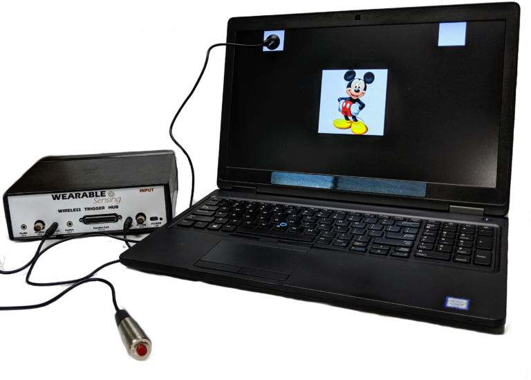
Our Wireless Trigger Hub simplifies the synchronization of DSI headsets with other devices. It features:
An additional benefit of the Trigger Hub design is that it allows synchronization across multiple data sources that are distributed across multiple systems, each of which running at its own clock rate. One such case commonly experienced in EEG experiments involves the synchronization of EEG and eye-tracking measurements, where the inevitable clock drift that arises between two systems during extended measurements creates difficulty in aligning data to events across the two systems.
The DSI-24 has 3 auxiliary inputs on the headset, which allows for automatic synchronization of Wearable Sensing’s auxiliary sensors to the EEG. The sensors available include ECG, EMG, EOG, GSR, RESP, & TEMP. The sensor data is collected and recorded in our data acquisition software, DSI-Streamer, where you can view the EEG and Aux sensors in real-time.
EEG Channels
Fp1, Fp2, Fz, F3, F4, F7, F8, Cz, C3, C4, T7/T3, T8/T4, Pz, P3, P4, P7/T5, P8/T6, O1, O2, A1, A2
Reference / Ground
Common Mode Follower / Fpz
Head Size Range
Adult Size: 52cm – 62cm circumference
Child Size: 48cm – 54cm circumference
Sampling Rate
300 Hz (600Hz upgrade available)
Bandwidth
0.003 – 150 Hz
A/D resolution
0.317 μV referred to input
Input Impedance (1Hz)
47 GΩ
CMRR
> 120 dB
Amplifier / Digitizer
16 bits / 24 channels
Wireless
Bluetooth
Wireless Range
10 m
Run-time
> 24 Hours, Hot-Swappable Batteries
Onboard Storage
~ 68 Hours (available option)
Data Acquisition
Real time, evoked potentials
Signal Quality Monitoring
Continuous impedance, Baseline offset, Noise (1-50 Hz)
Data Type
Raw and Filtered Data available
File Type
.CSV and .EDF
Data Output Streaming
TCP/IP socket, API (C Based), LSL
Cognitive State Classification
Brain Computer Interface
SSVEP BCI Algorithms; BCI2000; OpenViBE; PsychoPy; BCILab
Data Integration / Analysis
CAPTIV; Lab Streaming Layer; NeuroPype; BrainStorm; NeuroVIS
Neurofeedback
Applied Neuroscience NeuroGuide; Brainmaster Brain Avatar; EEGer
Neuromarketing
CAPTIV Neurolab
Presentation
Presentation; E-Prime
Schneefeld, F; Doelling, K; Marchesotti, S; Schwartz, S; Igloi, K; Giraud, AL; Arnal, LH
Salient 40 Hz sounds probe affective aversion and neural excitability Journal Article
In: bioRxiv, 2022.
@article{schneefeld2022salient,

title = {Salient 40 Hz sounds probe affective aversion and neural excitability},

author = {F Schneefeld and K Doelling and S Marchesotti and S Schwartz and K Igloi and AL Giraud and LH Arnal},

doi = {https://doi.org/10.1101/2022.02.26.482077},

year  = {2022},

date = {2022-03-01},

urldate = {2022-01-01},

journal = {bioRxiv},

publisher = {Cold Spring Harbor Laboratory},

abstract = {The human auditory system is not equally reactive to all frequencies of the audible spectrum. Emotional and behavioral reactions to loud or aversive acoustic features can vary from one individual to another, to the point that some exhibit exaggerated or even pathological responses to certain sounds. The neural mechanisms underlying these interindividual differences remain unclear. Whether distinct aversion profiles map onto neural excitability at the individual level needs to be tested. Here, we measured behavioral and EEG responses to click trains (from 10 to 250 Hz, spanning the roughness and pitch perceptual ranges) to test the hypothesis that interindividual variability in aversion to rough sounds is reflected in neural response differences between participants. Linking subjective aversion to 40 Hz steady-state EEG responses, we demonstrate that participants experiencing enhanced aversion to roughness also show stronger neural responses to this attribute. Interestingly, this pattern also correlates with inter-individual anxiety levels, suggesting that this personality trait might interact with subjective sensitivity and neural excitability to these sounds. These results support the idea that 40 Hz sounds can probe the excitability of non-canonical auditory systems involved in exogenous salience processing and aversive responses at the individual level. By linking subjective aversion to neural excitability, 40 Hz sounds provide neuromarkers relevant to a variety of pathological conditions, such as those featuring enhanced emotional sensitivity (hyperacusis, anxiety) or aberrant neural responses at 40 Hz (autism, schizophrenia).},

keywords = {},

pubstate = {published},

tppubtype = {article}

}
Haro, Stephanie; Rao, Hrishikesh M; Quatieri, Thomas F; Smalt, Christopher J
EEG alpha and pupil diameter reflect endogenous auditory attention switching and listening effort Journal Article
In: European Journal of Neuroscience, vol. 55, no. 5, pp. 1262–1277, 2022.
@article{haro2022eeg,

title = {EEG alpha and pupil diameter reflect endogenous auditory attention switching and listening effort},

author = {Stephanie Haro and Hrishikesh M Rao and Thomas F Quatieri and Christopher J Smalt},

doi = {https://doi.org/10.1111/ejn.15616},

year  = {2022},

date = {2022-01-30},

urldate = {2022-01-01},

journal = {European Journal of Neuroscience},

volume = {55},

number = {5},

pages = {1262--1277},

abstract = {Everyday environments often contain distracting competing talkers and background noise, requiring listeners to focus their attention on one acoustic source and reject others. During this auditory attention task, listeners may naturally interrupt their sustained attention and switch attended sources. The effort required to perform this attention switch has not been well studied in the context of competing continuous speech. In this work, we developed two variants of endogenous attention switching and a sustained attention control. We characterized these three experimental conditions under the context of decoding auditory attention, while simultaneously evaluating listening effort and neural markers of spatial-audio cues. A least-squares, electroencephalography (EEG)-based, attention decoding algorithm was implemented across all conditions. It achieved an accuracy of 69.4% and 64.0% when computed over nonoverlapping 10 and 5-s correlation windows, respectively. Both decoders illustrated smooth transitions in the attended talker prediction through switches at approximately half of the analysis window size (e.g., the mean lag taken across the two switch conditions was 2.2 s when the 5-s correlation window was used). Expended listening effort, as measured by simultaneous EEG and pupillometry, was also a strong indicator of whether the listeners sustained attention or performed an endogenous attention switch (peak pupil diameter measure [ ] and minimum parietal alpha power measure [ ]). We additionally found evidence of talker spatial cues in the form of centrotemporal alpha power lateralization ( ). These results suggest that listener effort and spatial cues may be promising features to pursue in a decoding context, in addition to speech-based features.},

keywords = {},

pubstate = {published},

tppubtype = {article}

}
Gubler, Dani`ele A; Zeiss, Stephan; Egloff, Niklaus; Frickmann, Frank; Goetze, Benjamin; Harnik, Michael; Streitberger, Konrad; Troche, Stefan J; others,
The effect of chronic pain on voluntary and involuntary capture of attention: An event-related potential study. Journal Article
In: Behavioral neuroscience, vol. 136, no. 2, pp. 195, 2022.
@article{gubler2022effect,

title = {The effect of chronic pain on voluntary and involuntary capture of attention: An event-related potential study.},

author = {Dani`ele A Gubler and Stephan Zeiss and Niklaus Egloff and Frank Frickmann and Benjamin Goetze and Michael Harnik and Konrad Streitberger and Stefan J Troche and others},

doi = {https://doi.org/10.1037/bne0000375},

year  = {2022},

date = {2022-01-01},

urldate = {2022-01-01},

journal = {Behavioral neuroscience},

volume = {136},

number = {2},

pages = {195},

publisher = {American Psychological Association},

abstract = {Although the interrupting effect of chronic pain on voluntary-directed attention is well-documented, research on the impact of chronic pain on involuntary-directed attention remains incomplete. This study aimed to investigate the influence of chronic pain on involuntary as well as voluntary allocation of attention as, respectively, indexed by the P3a and P3b components in the event-related potential derived from the electroencephalogram. Both involuntary and voluntary captures of attention were compared between 33 patients with chronic pain and 33 healthy controls using an auditory three-stimulus oddball task (with standard, target, and unexpected distractor tones). The results revealed a reduced P3a amplitude as well as a reduced P3b amplitude in patients with chronic pain compared to healthy controls, indicating a detrimental effect of chronic pain on involuntary and voluntary attention, respectively. This study extends the picture of the impairing effects of chronic pain on attentional allocation to a current task and attentional allocation to information outside the focus of attention. (PsycInfo Database Record (c) 2022 APA, all rights reserved)},

keywords = {},

pubstate = {published},

tppubtype = {article}

}
Arakaki, Xianghong; Hung, Shao-Min; Rochart, Roger; Fonteh, Alfred N; Harrington, Michael G
Alpha desynchronization during Stroop test unmasks cognitively healthy individuals with abnormal CSF Amyloid/Tau Journal Article
In: Neurobiology of Aging, 2021.
@article{arakaki2021alpha,

title = {Alpha desynchronization during Stroop test unmasks cognitively healthy individuals with abnormal CSF Amyloid/Tau},

author = {Xianghong Arakaki and Shao-Min Hung and Roger Rochart and Alfred N Fonteh and Michael G Harrington},

doi = {https://doi.org/10.1016/j.neurobiolaging.2021.11.009},

year  = {2021},

date = {2021-12-05},

journal = {Neurobiology of Aging},

publisher = {Elsevier},

abstract = {Synaptic dysfunctions precede cognitive decline in Alzheimer's disease (AD) by decades, affect executive functions, and can be detected by quantitative electroencephalography (qEEG). We used qEEG combined with Stroop testing to identify changes of inhibitory controls in cognitively healthy individuals with an abnormal versus normal ratio of cerebrospinal fluid (CSF) amyloid/total-tau. We studied two groups of participants (60-94 years) with either normal (CH-NAT or controls, n = 20) or abnormal (CH-PAT, n = 21) CSF amyloid/tau ratio. We compared: alpha event-related desynchronization (ERD), alpha spectral entropy (SE), and their relationships with estimated cognitive reserve. CH-PATs had more negative occipital alpha ERD, and higher frontal and occipital alpha SE during low load congruent trials, indicating hyperactivity. CH-PATs demonstrated fewer frontal SE changes with higher load, incongruent Stroop testing. Correlations of alpha ERD with estimated cognitive reserve were significant in CH-PATs but not in CH-NATs. These results suggested compensatory hyperactivity in CH-PATs compared to CH-NATs. We did not find differences in alpha ERD comparisons with individual CSF amyloid(A), p-tau(T), total-tau(N) biomarkers.},

keywords = {},

pubstate = {published},

tppubtype = {article}

}
Dong, Sunghee; Jin, Yan; Bak, SuJin; Yoon, Bumchul; Jeong, Jichai
Explainable Convolutional Neural Network to Investigate Age-Related Changes in Multi-Order Functional Connectivity Journal Article
In: Electronics, vol. 10, no. 23, pp. 3020, 2021.
@article{dong2021explainable,

title = {Explainable Convolutional Neural Network to Investigate Age-Related Changes in Multi-Order Functional Connectivity},

author = {Sunghee Dong and Yan Jin and SuJin Bak and Bumchul Yoon and Jichai Jeong},

doi = {https://doi.org/10.3390/electronics10233020},

year  = {2021},

date = {2021-12-03},

journal = {Electronics},

volume = {10},

number = {23},

pages = {3020},

publisher = {Multidisciplinary Digital Publishing Institute},

abstract = {Functional connectivity (FC) is a potential candidate that can increase the performance of brain-computer interfaces (BCIs) in the elderly because of its compensatory role in neural circuits. However, it is difficult to decode FC by the current machine learning techniques because of a lack of physiological understanding. To investigate the suitability of FC in BCIs for the elderly, we propose the decoding of lower- and higher-order FC using a convolutional neural network (CNN) in six cognitive-motor tasks. The layer-wise relevance propagation (LRP) method describes how age-related changes in FCs impact BCI applications for the elderly compared to younger adults. A total of 17 young adults (24.5±2.7 years) and 12 older (72.5±3.2 years) adults were recruited to perform tasks related to hand-force control with or without mental calculation. The CNN yielded a six-class classification accuracy of 75.3% in the elderly, exceeding the 70.7% accuracy for the younger adults. In the elderly, the proposed method increased the classification accuracy by 88.3% compared to the filter-bank common spatial pattern. The LRP results revealed that both lower- and higher-order FCs were dominantly overactivated in the prefrontal lobe, depending on the task type. These findings suggest a promising application of multi-order FC with deep learning on BCI systems for the elderly},

keywords = {},

pubstate = {published},

tppubtype = {article}

}
Arechavala, Rebecca Johnson; Rochart, Roger; Kloner, Robert A; Liu, Anqi; Wu, Daw-An; Hung, Shao-Min; Shimojo, Shinsuke; Fonteh, Alfred N; Kleinman, Michael T; Harrington, Michael G; others,
In: International Journal of Psychophysiology, vol. 170, pp. 102–111, 2021.
@article{arechavala2021task,

title = {Task switching reveals abnormal brain-heart electrophysiological signatures in cognitively healthy individuals with abnormal CSF amyloid/tau, a pilot study},

author = {Rebecca Johnson Arechavala and Roger Rochart and Robert A Kloner and Anqi Liu and Daw-An Wu and Shao-Min Hung and Shinsuke Shimojo and Alfred N Fonteh and Michael T Kleinman and Michael G Harrington and others},

doi = {https://doi.org/10.1016/j.ijpsycho.2021.10.007},

year  = {2021},

date = {2021-12-01},

journal = {International Journal of Psychophysiology},

volume = {170},

pages = {102--111},

publisher = {Elsevier},

abstract = {Electroencephalographic (EEG) alpha oscillations have been related to heart rate variability (HRV) and both change in Alzheimer's disease (AD). We explored if task switching reveals altered alpha power and HRV in cognitively healthy individuals with AD pathology in cerebrospinal fluid (CSF) and whether HRV improves the AD pathology classification by alpha power alone.


We compared low and high alpha event-related desynchronization (ERD) and HRV parameters during task switch testing between two groups of cognitively healthy participants classified by CSF amyloid/tau ratio: normal (CH-NAT, n = 19) or pathological (CH-PAT, n = 27). For the task switching paradigm, participants were required to name the color or word for each colored word stimulus, with two sequential stimuli per trial. Trials include color (cC) or word (wW) repeats with low load repeating, and word (cW) or color switch (wC) for high load switching. HRV was assessed for RR interval, standard deviation of RR-intervals (SDNN) and root mean squared successive differences (RMSSD) in time domain, and low frequency (LF), high frequency (HF), and LF/HF ratio in frequency domain.


Results showed that CH-PATs compared to CH-NATs presented: 1) increased (less negative) low alpha ERD during low load repeat trials and lower word switch cost (low alpha: p = 0.008, Cohen's d = −0.83, 95% confidence interval −1.44 to −0.22, and high alpha: p = 0.019, Cohen's d = −0.73, 95% confidence interval −1.34 to −0.13); 2) decreasing HRV from rest to task, suggesting hyper-activated sympatho-vagal responses. 3) CH-PATs classification by alpha ERD was improved by supplementing HRV signatures, supporting a potentially compromised brain-heart interoceptive regulation in CH-PATs. Further experiments are needed to validate these findings for clinical significance.},

keywords = {},

pubstate = {published},

tppubtype = {article}

}
Jung, Mijung; Lee, Mikyoung
The Effect of a Mindfulness-Based Education Program on Brain Waves and the Autonomic Nervous System in University Students Journal Article
In: Healthcare 2021, vol. 9, no. 11, pp. 1606, 2021.
@article{jung2021effect,

title = {The Effect of a Mindfulness-Based Education Program on Brain Waves and the Autonomic Nervous System in University Students},

author = {Mijung Jung and Mikyoung Lee},

doi = {https://doi.org/10.3390/healthcare9111606},

year  = {2021},

date = {2021-11-22},

booktitle = {Healthcare},

journal = {Healthcare 2021},

volume = {9},

number = {11},

pages = {1606},

organization = {Multidisciplinary Digital Publishing Institute},

abstract = {Background: Mindfulness, defined as the awareness emerging from purposefully paying attention to the present moment, has been shown to be effective in reducing stress and, thus, promoting psychological well-being. This study investigated the effects of a mindfulness-based education program on mindfulness, brain waves, and the autonomic nervous system (ANS) in university students in Korea. Methods: This study is a quantitative and experimental research with a single-group pre-post design. Six sessions of mindfulness-based intervention were applied. In total, 42 students completed a mindfulness questionnaire before and after the intervention, and 28 among them completed pre-intervention and post-intervention measures of brain waves and ANS. Results: The level of mindfulness increased in the participants after intervention. Regarding brain waves, the alpha and theta waves increased, but the beta waves decreased. There was no significant difference in the ANS, presenting no change in heart rate variability. Conclusions: We identified the positive effects of the mindfulness-based education program for university students. The findings indicate that this program may help students not only relax, but also generate a mindfulness state in stressful situations, potentially leading to a successful university life. This study can be used as a basis for quality improvement and sustainability of mindfulness-based education programs for university students.},

keywords = {},

pubstate = {published},

tppubtype = {article}

}
Seo, Ssang-Hee
Derivation of EEG Spectrum-based Feature Parameters for Mental Fatigue Determination Journal Article
In: Journal of Convergence for Information Technology, vol. 11, no. 10, pp. 10–19, 2021.
@article{seo2021derivation,

title = {Derivation of EEG Spectrum-based Feature Parameters for Mental Fatigue Determination},

author = {Ssang-Hee Seo},

doi = {https://doi.org/10.22156/CS4SMB.2021.11.10.010},

year  = {2021},

date = {2021-10-28},

journal = {Journal of Convergence for Information Technology},

volume = {11},

number = {10},

pages = {10--19},

publisher = {Convergence Society for SMB},

abstract = {In this paper, we tried to derive characteristic parameters that reflect mental fatigue through EEG measurement and analysis. For this purpose, mental fatigue was induced through a resting state with eyes closed and performing subtraction operations in mental arithmetic for 30 minutes. Five subjects participated in the experiment, and all subjects were right-handed male students in university, with an average age of 25.5 years. Spectral analysis was performed on the EEG collected at the beginning and the end of the experiment to derive feature parameters reflecting mental fatigue. As a result of the analysis, the absolute power of the alpha band in the occipital lobe and the temporal lobe increased as the mental fatigue increased, while the relative power decreased. Also, the difference in power between resting state and task state showed that the relative power was larger than the absolute power. These results indicate that alpha relative power in the occipital lobe and temporal lobe is a feature parameter reflecting mental fatigue. The results of this study can be utilized as feature parameters for the development of an automated system for mental fatigue determination such as fatigue and drowsiness while driving.},

keywords = {},

pubstate = {published},

tppubtype = {article}

}
Chakravarty, Sumit; Xie, Ying; Le, Linh; Johnson, John; Hales, Michael
Comparison Between Active and Passive Attention Using EEG Waves and Deep Neural Network Conference
International Conference on Brain Informatics, vol. 12960, Springer 2021, ISBN: 978-3-030-86993-9.
@conference{chakravarty2021comparison,

title = {Comparison Between Active and Passive Attention Using EEG Waves and Deep Neural Network},

author = {Sumit Chakravarty and Ying Xie and Linh Le and John Johnson and Michael Hales},

doi = {https://doi.org/10.1007/978-3-030-86993-9_27},

isbn = {978-3-030-86993-9},

year  = {2021},

date = {2021-09-15},

booktitle = {International Conference on Brain Informatics},

volume = {12960},

pages = {287--298},

organization = {Springer},

abstract = {A person’s state of attentiveness can be affected by various outside factors. Having energy, feeling tired, or even simply being distracted all play a role in someone’s level of attention. The task at hand can potentially affect the person’s attention or concentration level as well. In terms of students who take online courses, constantly watching lectures and conducting these courses solely online can cause lack of concentration or attention. Attention can be considered in two categories: passive or active. Conducting active and passive attention-based trials can reveal different states of attentiveness. This paper compares active and passive attention trial results of the two states, wide awake and tired. This has been done in order to uncover a difference in results between the two states. The data analyzed throughout this paper was collected from DSI 24 EEG equipment, and the generated EEG is processed through a 3D Convolutional Neural Network (CNN) to produce results. Three passive attention trials and three active attention trials were performed on seven subjects, while they were wide awake and when they were tired. The experiments on the preprocessed data results in accuracies as high as 81.78% for passive attention detection accuracy and 63.67% for active attention detection accuracy, which shown a clear ability to separate between the two attention categories.},

keywords = {},

pubstate = {published},

tppubtype = {conference}

}
Attar, Eyad Talal; Balasubramanian, Vignesh; Subasi, Ersoy; Kaya, Mehmet
Stress analysis based on simultaneous heart rate variability and EEG monitoring Journal Article
In: IEEE Journal of Translational Engineering in Health and Medicine, vol. 9, pp. 1–7, 2021.
@article{attar2021stress,

title = {Stress analysis based on simultaneous heart rate variability and EEG monitoring},

author = {Eyad Talal Attar and Vignesh Balasubramanian and Ersoy Subasi and Mehmet Kaya},

doi = {https://doi.org/10.1109/JTEHM.2021.3106803},

year  = {2021},

date = {2021-08-23},

urldate = {2021-01-01},

journal = {IEEE Journal of Translational Engineering in Health and Medicine},

volume = {9},

pages = {1–7},

publisher = {IEEE},

abstract = {Objective: Stress is a significant risk factor for various diseases such as hypertension, heart attack, stroke, and even sudden death. Stress can also lead to psychological and behavioral disorders. Heart rate variability (HRV) can reflect changes in stress levels while other physiological factors, like blood pressure, are within acceptable ranges. Electroencephalogram (EEG) is a vital technique for studying brain activities and provides useful data regarding changes in mental status. This study incorporates EEG and a detailed HRV analysis to have a better understanding and analysis of stress. Investigating the correlation between EEG and HRV under stress conditions is valuable since they provide complementary information regarding stress. Methods: Simultaneous electrocardiogram (ECG) and EEG recordings were obtained from fifteen subjects. HRV /EEG features were analyzed and compared in rest, stress, and meditation conditions. A one-way ANOVA and correlation coefficient were used for statistical analysis to explore the correlation between HRV features and features extracted from EEG. Results: The HRV features LF (low frequency), HF (high frequency), LF/HF, and rMSSD (root mean square of the successive differences) correlated with EEG features, including alpha power band in the left hemisphere and alpha band power asymmetry. Conclusion: This study demonstrated five significant relationships between EEG and HRV features associated with stress. The ability to use stress-related EEG features in combination with correlated HRV features could help improve detecting stress and monitoring the progress of stress treatments/therapies. The outcomes of this study could enhance the efficiency of stress management technologies such as meditation studies and bio-feedback training.},

keywords = {},

pubstate = {published},

tppubtype = {article}

}
Please fill out the form and provide a brief description of your application so we can help match you with products that will meet your specific needs.
Please fill out the form and provide a brief description of your application so we can help match you with products that will meet your specific needs.