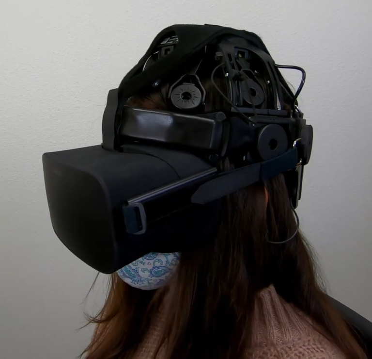
Wearable Sensing’s wireless DSI-24 is the leading dry electrode EEG system in terms of signal quality and comfort. The DSI-24 takes on average less than 3 minutes to set up, making it the ideal solution for scientists in need of a simple, easy to use, EEG system. Our patented sensor technology not only delivers uncompromised signal quality but also enables our system to be virtually immune against motion and electrical artifacts. As a result, the DSI-24 can be utilized in virtual or augmented reality, while also allowing researchers to take their experiments out of the lab, and into the real world.
The DSI-24 has sensors that provide full head coverage with 19 electrodes on the head, 2 earclip sensors, and also has 3 built-in auxiliary inputs for acquisition of up to 3 auxiliary sensors. It also has an 8-bit trigger input to synchronize with other devices such as Eye-Tracking, Motion (IMU), and more.
Used around the world by leaders in Research, Neurofeedback, Neuromarketing, Brain-Computer Interfaces, & Neuroergonomics.
With over 90% correlation to research-grade wet EEG systems, the dry sensor interface (DSI) offers unparalleled quality and performance
Multiple adjustment points and a foam pad lined interior enable the system to be worn for up to 8 hours on any head shape or size
All DSI systems include free, unlimited licenses of DSI-Streamer, our data acquisition software which can record raw data, in .csv and .edf file formats
Faraday cage's, spring-loaded electrodes, and our patented common-mode follower technology, provides near immunity against electrical and motion artifacts
Using 70% isopropyl alcohol and a cleaning brush, the DSI-24 only takes a minute to clean, 3 minutes to dry, and can be up and running on the next subject in minutes
All DSI systems include our free C based .dll API, which enables users to pull the raw data directly from the headset, for custom software on Windows, Mac OS, Linux, and ARM
The DSI-24 was designed for ultra-rapid setup, taking on average less than 3 minutes to don, and works on any type of hair, including long hair, thick hair, afros, and more
DSI headsets have active sensors, amplifiers, digitizers, batteries, onboard storage, and wireless transmission, making them complete, mobile, wearable EEG systems
DSI systems exclusively work with QStates, a machine learning algorithm for cognitive classification on states such as mental workload, engagement, and fatigue
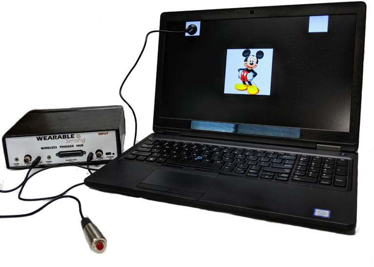
Our Wireless Trigger Hub simplifies the synchronization of DSI headsets with other devices. It features:
An additional benefit of the Trigger Hub design is that it allows synchronization across multiple data sources that are distributed across multiple systems, each of which running at its own clock rate. One such case commonly experienced in EEG experiments involves the synchronization of EEG and eye-tracking measurements, where the inevitable clock drift that arises between two systems during extended measurements creates difficulty in aligning data to events across the two systems.
The DSI-24 has 3 auxiliary inputs on the headset, which allows for automatic synchronization of Wearable Sensing’s auxiliary sensors to the EEG. The sensors available include ECG, EMG, EOG, GSR, RESP, & TEMP. The sensor data is collected and recorded in our data acquisition software, DSI-Streamer, where you can view the EEG and Aux sensors in real-time.
EEG Channels
Fp1, Fp2, Fz, F3, F4, F7, F8, Cz, C3, C4, T7/T3, T8/T4, Pz, P3, P4, P7/T5, P8/T6, O1, O2, A1, A2
Reference / Ground
Common Mode Follower / Fpz
Head Size Range
Adult Size: 52cm – 62cm circumference
Child Size: 48cm – 54cm circumference
Sampling Rate
300 Hz (600Hz upgrade available)
Bandwidth
0.003 – 150 Hz
A/D resolution
0.317 μV referred to input
Input Impedance (1Hz)
47 GΩ
CMRR
> 120 dB
Amplifier / Digitizer
16 bits / 24 channels
Wireless
Bluetooth
Wireless Range
10 m
Run-time
> 24 Hours, Hot-Swappable Batteries
Onboard Storage
~ 68 Hours (available option)
Data Acquisition
Real time, evoked potentials
Signal Quality Monitoring
Continuous impedance, Baseline offset, Noise (1-50 Hz)
Data Type
Raw and Filtered Data available
File Type
.CSV and .EDF
Data Output Streaming
TCP/IP socket, API (C Based), LSL
Cognitive State Classification
Brain Computer Interface
SSVEP BCI Algorithms; BCI2000; OpenViBE; PsychoPy; BCILab
Data Integration / Analysis
CAPTIV; Lab Streaming Layer; NeuroPype; BrainStorm; NeuroVIS
Neurofeedback
Applied Neuroscience NeuroGuide; Brainmaster Brain Avatar; EEGer
Neuromarketing
CAPTIV Neurolab
Presentation
Presentation; E-Prime
Seo, Ssang-Hee
Derivation of EEG Spectrum-based Feature Parameters for Mental Fatigue Determination Journal Article
In: Journal of Convergence for Information Technology, vol. 11, no. 10, pp. 10–19, 2021.
@article{seo2021derivation,

title = {Derivation of EEG Spectrum-based Feature Parameters for Mental Fatigue Determination},

author = {Ssang-Hee Seo},

doi = {https://doi.org/10.22156/CS4SMB.2021.11.10.010},

year  = {2021},

date = {2021-10-28},

journal = {Journal of Convergence for Information Technology},

volume = {11},

number = {10},

pages = {10--19},

publisher = {Convergence Society for SMB},

abstract = {In this paper, we tried to derive characteristic parameters that reflect mental fatigue through EEG measurement and analysis. For this purpose, mental fatigue was induced through a resting state with eyes closed and performing subtraction operations in mental arithmetic for 30 minutes. Five subjects participated in the experiment, and all subjects were right-handed male students in university, with an average age of 25.5 years. Spectral analysis was performed on the EEG collected at the beginning and the end of the experiment to derive feature parameters reflecting mental fatigue. As a result of the analysis, the absolute power of the alpha band in the occipital lobe and the temporal lobe increased as the mental fatigue increased, while the relative power decreased. Also, the difference in power between resting state and task state showed that the relative power was larger than the absolute power. These results indicate that alpha relative power in the occipital lobe and temporal lobe is a feature parameter reflecting mental fatigue. The results of this study can be utilized as feature parameters for the development of an automated system for mental fatigue determination such as fatigue and drowsiness while driving.},

keywords = {},

pubstate = {published},

tppubtype = {article}

}
Chakravarty, Sumit; Xie, Ying; Le, Linh; Johnson, John; Hales, Michael
Comparison Between Active and Passive Attention Using EEG Waves and Deep Neural Network Conference
International Conference on Brain Informatics, vol. 12960, Springer 2021, ISBN: 978-3-030-86993-9.
@conference{chakravarty2021comparison,

title = {Comparison Between Active and Passive Attention Using EEG Waves and Deep Neural Network},

author = {Sumit Chakravarty and Ying Xie and Linh Le and John Johnson and Michael Hales},

doi = {https://doi.org/10.1007/978-3-030-86993-9_27},

isbn = {978-3-030-86993-9},

year  = {2021},

date = {2021-09-15},

booktitle = {International Conference on Brain Informatics},

volume = {12960},

pages = {287--298},

organization = {Springer},

abstract = {A person’s state of attentiveness can be affected by various outside factors. Having energy, feeling tired, or even simply being distracted all play a role in someone’s level of attention. The task at hand can potentially affect the person’s attention or concentration level as well. In terms of students who take online courses, constantly watching lectures and conducting these courses solely online can cause lack of concentration or attention. Attention can be considered in two categories: passive or active. Conducting active and passive attention-based trials can reveal different states of attentiveness. This paper compares active and passive attention trial results of the two states, wide awake and tired. This has been done in order to uncover a difference in results between the two states. The data analyzed throughout this paper was collected from DSI 24 EEG equipment, and the generated EEG is processed through a 3D Convolutional Neural Network (CNN) to produce results. Three passive attention trials and three active attention trials were performed on seven subjects, while they were wide awake and when they were tired. The experiments on the preprocessed data results in accuracies as high as 81.78% for passive attention detection accuracy and 63.67% for active attention detection accuracy, which shown a clear ability to separate between the two attention categories.},

keywords = {},

pubstate = {published},

tppubtype = {conference}

}
Lim, Hyunmi; Ku, Jeonghun
Superior Facilitation of an Action Observation Network by Congruent Character Movements in Brain--Computer Interface Action-Observation Games Journal Article
In: Cyberpsychology, Behavior, and Social Networking, vol. 24, no. 8, pp. 566–572, 2021.
@article{lim2021superior,

title = {Superior Facilitation of an Action Observation Network by Congruent Character Movements in Brain--Computer Interface Action-Observation Games},

author = {Hyunmi Lim and Jeonghun Ku},

doi = {https://doi.org/10.1089/cyber.2020.0231},

year  = {2021},

date = {2021-08-04},

journal = {Cyberpsychology, Behavior, and Social Networking},

volume = {24},

number = {8},

pages = {566--572},

publisher = {Mary Ann Liebert, Inc., publishers 140 Huguenot Street, 3rd Floor New~…},

abstract = {Action observation (AO) is a promising strategy for promoting motor function in neural rehabilitation. Recently, brain–computer interface (BCI)-AO game rehabilitation, which combines AO therapy with BCI technology, has been introduced to improve the effectiveness of rehabilitation. This approach can improve motor learning by providing feedback, which can be interactive in an observation task, and the game contents of the BCI-AO game paradigm can affect rehabilitation. In this study, the effects of congruent rather than incongruent feedback in a BCI-AO game on mirror neurons were investigated. Specifically, the mu suppression with congruent and incongruent BCI-AO games was measured in 17 healthy adults. The mu suppression in the central motor cortex was significantly higher with the congruent BCI-AO game than with the incongruent one. In addition, the satisfaction evaluation results were excellent for the congruent case. These results support the fact that providing feedback congruent with the motion of an action video facilitates mirror neuron activity and can offer useful guidelines for the design of BCI-AO games for rehabilitation},

keywords = {},

pubstate = {published},

tppubtype = {article}

}
Lin, Chun-Ling; Hsieh, Ya-Wen; Chen, Hui-Ya
Age-related differences in alpha and beta band activity in sensory association brain areas during challenging sensory tasks Journal Article
In: Behavioural Brain Research, vol. 408, pp. 113279, 2021.
@article{lin2021age,

title = {Age-related differences in alpha and beta band activity in sensory association brain areas during challenging sensory tasks},

author = {Chun-Ling Lin and Ya-Wen Hsieh and Hui-Ya Chen},

doi = {https://doi.org/10.1016/j.bbr.2021.113279},

year  = {2021},

date = {2021-06-25},

journal = {Behavioural Brain Research},

volume = {408},

pages = {113279},

publisher = {Elsevier},

abstract = {Sensory challenges to postural balance are daily threats for elderly individuals. This study examined electroencephalography (EEG) in alpha and beta bands in sensory association areas during the Sensory Organization Test, involving withdrawal of visual or presenting misleading somatosensory inputs, in twelve young and twelve elderly participants. The results showed stepwise deterioration in behavioral performance in four conditions, with group effects that were amplified with combined sensory challenges. With eye closure, alpha and beta activities increased in all sensory association areas. Fast beta activity increased in the bilateral parietal-temporal-occipital areas. Misleading somatosensory information effects on EEG activity were of smaller amplitude than eye closure effects and in a different direction. Decreased alpha activity in left parietal-temporal-occipital areas and decreased beta and fast beta activities in bilateral parietal-temporal-occipital areas were significant. Elderly participants had increased fast beta activity in the left temporal-occipital and bilateral occipital areas, indicative of sustained efforts that they made in all sensory conditions. Similar to the young participants, elderly participants with eyes closed showed increased alpha activity, although to a smaller degree, in bilateral temporal-occipital and left occipital areas. This might indicate a lack of efficacy in redistributing relative sensory weights when elderly participants dealt with eye closure. In summary, EEG power changes did not match the stepwise deterioration in behavioral data, but reflected different sensory strategies adopted by young and elderly participants to cope with eye closure or misleading somatosensory information based on the efficacy of these different strategies.},

keywords = {},

pubstate = {published},

tppubtype = {article}

}
Fan, Chen-Chen; Xie, Haiqun; Peng, Liang; Yang, Hongjun; Ni, Zhen-Liang; Wang, Guanán; Zhou, Yan-Jie; Chen, Sheng; Fang, Zhijie; Huang, Shuyun; others,
Group Feature Learning and Domain Adversarial Neural Network for aMCI Diagnosis System Based on EEG Conference
2021 International Conference on Robotics and Automation, 2021.
@conference{fan2021group,

title = {Group Feature Learning and Domain Adversarial Neural Network for aMCI Diagnosis System Based on EEG},

author = {Chen-Chen Fan and Haiqun Xie and Liang Peng and Hongjun Yang and Zhen-Liang Ni and Guanán Wang and Yan-Jie Zhou and Sheng Chen and Zhijie Fang and Shuyun Huang and others},

url = {https://arxiv.org/abs/2105.06270},

year  = {2021},

date = {2021-04-28},

booktitle = {2021 International Conference on Robotics and Automation},

journal = {2021 International Conference on Robotics and Automation},

abstract = {Medical diagnostic robot systems have been paid more and more attention due to its objectivity and accuracy. The diagnosis of mild cognitive impairment (MCI) is considered an effective means to prevent Alzheimer's disease (AD). Doctors diagnose MCI based on various clinical examinations, which are expensive and the diagnosis results rely on the knowledge of doctors. Therefore, it is necessary to develop a robot diagnostic system to eliminate the influence of human factors and obtain a higher accuracy rate. In this paper, we propose a novel Group Feature Domain Adversarial Neural Network (GF-DANN) for amnestic MCI (aMCI) diagnosis, which involves two important modules. A Group Feature Extraction (GFE) module is proposed to reduce individual differences by learning group-level features through adversarial learning. A Dual Branch Domain Adaptation (DBDA) module is carefully designed to reduce the distribution difference between the source and target domain in a domain adaption way. On three types of data set, GF-DANN achieves the best accuracy compared with classic machine learning and deep learning methods. On the DMS data set, GF-DANN has obtained an accuracy rate of 89.47%, and the sensitivity and specificity are 90% and 89%. In addition, by comparing three EEG data collection paradigms, our results demonstrate that the DMS paradigm has the potential to build an aMCI diagnose robot system.},

keywords = {},

pubstate = {published},

tppubtype = {conference}

}
Rustamov, Nabi; Sharma, Lokesh; Chiang, Sarah N; Burk, Carrie; Haroutounian, Simon; Leuthardt, Eric C
Spatial and frequency-specific electrophysiological signatures of tonic pain recovery in humans Journal Article
In: Neuroscience, vol. 465, pp. 23–37, 2021.
@article{rustamov2021spatial,

title = {Spatial and frequency-specific electrophysiological signatures of tonic pain recovery in humans},

author = {Nabi Rustamov and Lokesh Sharma and Sarah N Chiang and Carrie Burk and Simon Haroutounian and Eric C Leuthardt},

doi = {https://doi.org/10.1016/j.neuroscience.2021.04.008},

year  = {2021},

date = {2021-04-21},

urldate = {2021-01-01},

journal = {Neuroscience},

volume = {465},

pages = {23–37},

publisher = {Elsevier},

abstract = {The objective of this study was to comprehensively investigate patterns of brain activities associated with pain recovery following experimental tonic pain in humans. Specific electrophysiological features of pain recovery may either be monitored or be modulated through neurofeedback (NF) as a novel chronic pain treatment. The cold pressor test was applied with simultaneous electroencephalogram (EEG) recording. EEG data were acquired, and analyzed to define: (1) EEG power topography patterns of pain recovery; (2) source generators of pain recovery at cortical level; (3) changes in functional connectivity associated with pain recovery; (4) features of phase-amplitude coupling (PAC) as it relates to pain recovery. The novel finding of this study is that recovery from pain was characterized by significant theta power rebound at the left fronto-central area. The sources of theta power over-recovery were located in the left dorsolateral prefrontal cortex (DLPFC), cingulate cortex, left insula and contralateral sensorimotor cortex. These effects were paralleled by theta band connectivity increase within hemispheres in a prefrontal–somatosensory network and interhemispherically between prefrontal and parietal areas. In addition, this study revealed significant reduction in PAC between theta/alpha and gamma oscillations during recovery period following tonic pain. These findings have largely been replicated across two identical sessions. Our study emphasizes the association between pain recovery and left lateral prefrontal theta power rebound, and significant over-recovery of functional connectivity in prefrontal-sensorimotor neural network synchronized at theta frequencies. These findings may provide basis for chronic pain treatment by modulating neural oscillations at theta frequencies in left prefrontal cortex.},

keywords = {},

pubstate = {published},

tppubtype = {article}

}
Kim, Sanghee; Park, Hyejin; Choo, Seungyeon
Effects of Changes to Architectural Elements on Human Relaxation-Arousal Responses: Based on VR and EEG Journal Article
In: International Journal of Environmental Research and Public Health, vol. 18, no. 8, pp. 4305, 2021.
@article{kim2021effects,

title = {Effects of Changes to Architectural Elements on Human Relaxation-Arousal Responses: Based on VR and EEG},

author = {Sanghee Kim and Hyejin Park and Seungyeon Choo},

doi = {https://doi.org/10.3390/ijerph18084305},

year  = {2021},

date = {2021-04-19},

journal = {International Journal of Environmental Research and Public Health},

volume = {18},

number = {8},

pages = {4305},

publisher = {Multidisciplinary Digital Publishing Institute},

abstract = {This study combines electroencephalogram (EEG) with virtual reality (VR) technologies to measure the EEG responses of users experiencing changes to architectural elements. We analyze the ratio of alpha to beta waves (RAB) indicators to determine the pre- and poststimulation changes. In our methodology, thirty-three females experience using private rooms in a postpartum care center participated in the experiment. Their brain waves are measured while they are experiencing the VR space of a private room in a postpartum care center. Three architectural elements (i.e., aspect ratio of space, ceiling height, and window ratio) are varied in the VR space. In addition, a self-report questionnaire is administered to examine whether the responses are consistent with the results of the EEG response analysis. As a result, statistically significant differences (p < 0.05) are observed in the changes in the RAB indicator values of the pre- and poststimulation EEG while the subjects are experiencing the VR space where the architectural elements are varied. That is, the effects of the changes to architectural elements on users’ relaxation-arousal responses are statistically verified. Notably, in all the RAB indicator values where significant differences are observed, the poststimulation RAB decreases in comparison to the prestimulus ratios, which is indicative of the arousal response. However, the arousal levels vary across the architectural elements, which implies it would be possible to find out the elements that could induce less arousal response using the proposed method. Moreover, following the experience in the VR space, certain lobes of the brain (F4 and P3 EEG channels) show statistically significant differences in the relaxation-arousal responses. Unlike previous studies, which measured users’ physiological responses to abstract and primordial spatial elements, this study extends the boundaries of the literature by applying the architectural elements applicable to design in practice},

keywords = {},

pubstate = {published},

tppubtype = {article}

}
Zhang, Dong
Brain-Controlled Robotic Arm Based on Adaptive FBCCA Conference
Human Brain and Artificial Intelligence: Second International Workshop, HBAI 2020, Held in Conjunction with IJCAI-PRICAI 2020, Yokohama, Japan, January 7, 2021, Revised Selected Papers, Springer Nature 2021.
@conference{zhang2021brain,

title = {Brain-Controlled Robotic Arm Based on Adaptive FBCCA},

author = {Dong Zhang},

url = {https://link.springer.com/chapter/10.1007/978-981-16-1288-6_7},

year  = {2021},

date = {2021-04-08},

booktitle = {Human Brain and Artificial Intelligence: Second International Workshop, HBAI 2020, Held in Conjunction with IJCAI-PRICAI 2020, Yokohama, Japan, January 7, 2021, Revised Selected Papers},

pages = {102},

organization = {Springer Nature},

abstract = {The SSVEP-BCI system usually uses a fixed calculation time and a static window stop method to decode the EEG signal, which reduces the efficiency of the system. In response to this problem, this paper uses an adaptive FBCCA algorithm, which uses Bayesian estimation to dynamically find the optimal data length for result prediction, adapts to the differences between different trials and different individuals, and effectively improves system operation effectiveness. At the same time, through this method, this paper constructs a brain-controlled robotic arm grasping life assistance system based on adaptive FBCCA. In this paper, we selected 20 subjects and conducted a total of 400 experiments. A large number of experiments have verified that the system is available and the average recognition success rate is 95.5%. This also proves that the system can be applied to actual scenarios. Help the handicapped to use the brain to control the mechanical arm to grab the needed items to assist in daily life and improve the quality of life. In the future, SSVEP’s adaptive FBCCA decoding algorithm can be combined with the motor imaging brain-computer interface decoding algorithm to build a corresponding system to help patients with upper or lower limb movement disorders caused by stroke diseases to recover, and reshape the brain and Control connection of limbs.},

keywords = {},

pubstate = {published},

tppubtype = {conference}

}
Lim, Hyunmi; Ku, Jeonghun
Effect of repetitive neurofeedback training on brain activation during hand exercise Conference
2021 9th International Winter Conference on Brain-Computer Interface (BCI), IEEE 2021, ISBN: 978-1-7281-8486-9.
@conference{lim2021effect,

title = {Effect of repetitive neurofeedback training on brain activation during hand exercise},

author = {Hyunmi Lim and Jeonghun Ku},

doi = {10.1109/BCI51272.2021.9385315},

isbn = {978-1-7281-8486-9},

year  = {2021},

date = {2021-04-05},

booktitle = {2021 9th International Winter Conference on Brain-Computer Interface (BCI)},

pages = {1--3},

organization = {IEEE},

abstract = {In this study, we examined that neurofeedback training encouraging to make mu suppression over the motor cortex would effect on the brain activation of the motor cortex while performing hand movements. To investigate the effects of training with neurofeedback, we analyzed the pattern changes of the amount of the mu suppression during real hand movements just after every neurofeedback training. The healthy subjects were trained to increase the motor cortex activity by using motor imagery, specifically maximizing the difference of the mu power between C3 and C4 during neurofeedback training. We have observed that the changes of the mu suppression over the motor cortex become stronger as the neurofeedback training was repeated. These findings further suggest that motor imagery training using neurofeedback can be applied to patients with stroke or chronic neurological disorders.},

keywords = {},

pubstate = {published},

tppubtype = {conference}

}
Eldeeb, Safaa; Susam, Busra T; Akcakaya, Murat; Conner, Caitlin M; White, Susan W; Mazefsky, Carla A
Trial by trial EEG based BCI for distress versus non distress classification in individuals with ASD Journal Article
In: Scientific Reports, vol. 11, no. 1, pp. 1–13, 2021.
@article{eldeeb2021trial,

title = {Trial by trial EEG based BCI for distress versus non distress classification in individuals with ASD},

author = {Safaa Eldeeb and Busra T Susam and Murat Akcakaya and Caitlin M Conner and Susan W White and Carla A Mazefsky},

url = {https://www.nature.com/articles/s41598-021-85362-8},

year  = {2021},

date = {2021-03-16},

journal = {Scientific Reports},

volume = {11},

number = {1},

pages = {1--13},

publisher = {Nature Publishing Group},

abstract = {Autism spectrum disorder (ASD) is a neurodevelopmental disorder that is often accompanied by impaired emotion regulation (ER). There has been increasing emphasis on developing evidence-based approaches to improve ER in ASD. Electroencephalography (EEG) has shown success in reducing ASD symptoms when used in neurofeedback-based interventions. Also, certain EEG components are associated with ER. Our overarching goal is to develop a technology that will use EEG to monitor real-time changes in ER and perform intervention based on these changes. As a first step, an EEG-based brain computer interface that is based on an Affective Posner task was developed to identify patterns associated with ER on a single trial basis, and EEG data collected from 21 individuals with ASD. Accordingly, our aim in this study is to investigate EEG features that could differentiate between distress and non-distress conditions. Specifically, we investigate if the EEG time-locked to the visual feedback presentation could be used to classify between WIN (non-distress) and LOSE (distress) conditions in a game with deception. Results showed that the extracted EEG features could differentiate between WIN and LOSE conditions (average accuracy of 81%), LOSE and rest-EEG conditions (average accuracy 94.8%), and WIN and rest-EEG conditions (average accuracy 94.9%).},

keywords = {},

pubstate = {published},

tppubtype = {article}

}

Please fill out the form and provide a brief description of your application so we can help match you with products that will meet your specific needs.
Please fill out the form and provide a brief description of your application so we can help match you with products that will meet your specific needs.
Please fill out the form and provide a brief description of your application so we can help match you with products that will meet your specific needs.