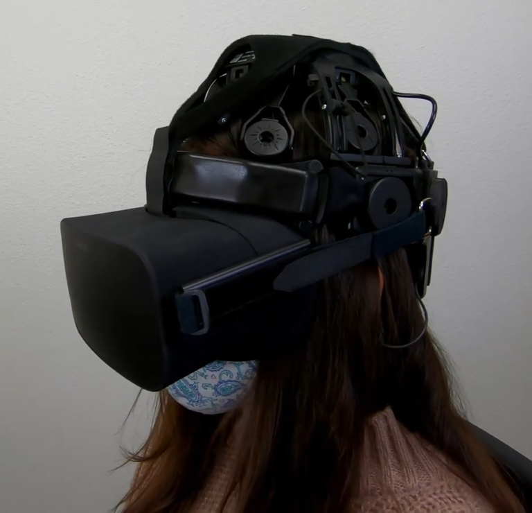
Wearable Sensing’s wireless DSI-24 is the leading dry electrode EEG system in terms of signal quality and comfort. The DSI-24 takes on average less than 3 minutes to set up, making it the ideal solution for scientists in need of a simple, easy to use, EEG system. Our patented sensor technology not only delivers uncompromised signal quality but also enables our system to be virtually immune against motion and electrical artifacts. As a result, the DSI-24 can be utilized in virtual or augmented reality, while also allowing researchers to take their experiments out of the lab, and into the real world.
The DSI-24 has sensors that provide full head coverage with 19 electrodes on the head, 2 earclip sensors, and also has 3 built-in auxiliary inputs for acquisition of up to 3 auxiliary sensors. It also has an 8-bit trigger input to synchronize with other devices such as Eye-Tracking, Motion (IMU), and more.
Used around the world by leaders in Research, Neurofeedback, Neuromarketing, Brain-Computer Interfaces, & Neuroergonomics.
With over 90% correlation to research-grade wet EEG systems, the dry sensor interface (DSI) offers unparalleled quality and performance
Multiple adjustment points and a foam pad lined interior enable the system to be worn for up to 8 hours on any head shape or size
All DSI systems include free, unlimited licenses of DSI-Streamer, our data acquisition software which can record raw data, in .csv and .edf file formats
Faraday cage's, spring-loaded electrodes, and our patented common-mode follower technology, provides near immunity against electrical and motion artifacts
Using 70% isopropyl alcohol and a cleaning brush, the DSI-24 only takes a minute to clean, 3 minutes to dry, and can be up and running on the next subject in minutes
All DSI systems include our free C based .dll API, which enables users to pull the raw data directly from the headset, for custom software on Windows, Mac OS, Linux, and ARM
The DSI-24 was designed for ultra-rapid setup, taking on average less than 3 minutes to don, and works on any type of hair, including long hair, thick hair, afros, and more
DSI headsets have active sensors, amplifiers, digitizers, batteries, onboard storage, and wireless transmission, making them complete, mobile, wearable EEG systems
DSI systems exclusively work with QStates, a machine learning algorithm for cognitive classification on states such as mental workload, engagement, and fatigue
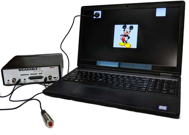
Our Wireless Trigger Hub simplifies the synchronization of DSI headsets with other devices. It features:
An additional benefit of the Trigger Hub design is that it allows synchronization across multiple data sources that are distributed across multiple systems, each of which running at its own clock rate. One such case commonly experienced in EEG experiments involves the synchronization of EEG and eye-tracking measurements, where the inevitable clock drift that arises between two systems during extended measurements creates difficulty in aligning data to events across the two systems.
The DSI-24 has 3 auxiliary inputs on the headset, which allows for automatic synchronization of Wearable Sensing’s auxiliary sensors to the EEG. The sensors available include ECG, EMG, EOG, GSR, RESP, & TEMP. The sensor data is collected and recorded in our data acquisition software, DSI-Streamer, where you can view the EEG and Aux sensors in real-time.
EEG Channels
Fp1, Fp2, Fz, F3, F4, F7, F8, Cz, C3, C4, T7/T3, T8/T4, Pz, P3, P4, P7/T5, P8/T6, O1, O2, A1, A2
Reference / Ground
Common Mode Follower / Fpz
Head Size Range
Adult Size: 52cm – 62cm circumference
Child Size: 48cm – 54cm circumference
Sampling Rate
300 Hz (600Hz upgrade available)
Bandwidth
0.003 – 150 Hz
A/D resolution
0.317 μV referred to input
Input Impedance (1Hz)
47 GΩ
CMRR
> 120 dB
Amplifier / Digitizer
16 bits / 24 channels
Wireless
Bluetooth
Wireless Range
10 m
Run-time
> 24 Hours, Hot-Swappable Batteries
Onboard Storage
~ 68 Hours (available option)
Data Acquisition
Real time, evoked potentials
Signal Quality Monitoring
Continuous impedance, Baseline offset, Noise (1-50 Hz)
Data Type
Raw and Filtered Data available
File Type
.CSV and .EDF
Data Output Streaming
TCP/IP socket, API (C Based), LSL
Cognitive State Classification
Brain Computer Interface
SSVEP BCI Algorithms; BCI2000; OpenViBE; PsychoPy; BCILab
Data Integration / Analysis
CAPTIV; Lab Streaming Layer; NeuroPype; BrainStorm; NeuroVIS
Neurofeedback
Applied Neuroscience NeuroGuide; Brainmaster Brain Avatar; EEGer
Neuromarketing
CAPTIV Neurolab
Presentation
Presentation; E-Prime
Haar Millo, S; Faisal, A
Brain Activity Reveals Multiple Motor-Learning Mechanisms in a Real-World Task Journal Article
In: Frontiers in Human Neuroscience, vol. 14, pp. 354, 2020, ISBN: 1662-5161.
@article{haarbrain,

title = {Brain Activity Reveals Multiple Motor-Learning Mechanisms in a Real-World Task},

author = {Haar Millo, S and Faisal, A},

doi = {10.3389/fnhum.2020.00354},

isbn = {1662-5161},

year  = {2020},

date = {2020-09-02},

journal = {Frontiers in Human Neuroscience},

volume = {14},

pages = {354},

publisher = {Frontiers Media},

abstract = {Many recent studies found signatures of motor learning in neural beta oscillations (13–30 Hz), and specifically in the post-movement beta rebound (PMBR). All these studies were in controlled laboratory-tasks in which the task designed to induce the studied learning mechanism. Interestingly, these studies reported opposing dynamics of the PMBR magnitude over learning for the error-based and reward-based tasks (increase vs. decrease, respectively). Here, we explored the PMBR dynamics during real-world motor-skill-learning in a billiards task using mobile-brain-imaging. Our EEG recordings highlight the opposing dynamics of PMBR magnitudes (increase vs. decrease) between different subjects performing the same task. The groups of subjects, defined by their neural dynamics, also showed behavioral differences expected for different learning mechanisms. Our results suggest that when faced with the complexity of the real-world different subjects might use different learning mechanisms for the same complex task. We speculate that all subjects combine multi-modal mechanisms of learning, but different subjects have different predominant learning mechanisms.},

keywords = {},

pubstate = {published},

tppubtype = {article}

}
Kim, Young-June; Park, Jin-Hong; Cho, Young-Suk; Kim, Keum-Sook
In: Journal of Convergence for Information Technology, vol. 10, no. 8, pp. 203–212, 2020.
@article{kim2020effect,

title = {The Effect of Cognitive Rehabilitation Program Using Virtual Reality (VR) Contents on Cognitive function, Depression, Upper Extremity Function and Activities of Daily Living in the Elderly},

author = {Young-June Kim and Jin-Hong Park and Young-Suk Cho and Keum-Sook Kim},

url = {https://www.koreascience.or.kr/article/JAKO202024852036461.page},

year  = {2020},

date = {2020-08-28},

journal = {Journal of Convergence for Information Technology},

volume = {10},

number = {8},

pages = {203--212},

publisher = {Convergence Society for SMB},

abstract = {The purpose of this study was to investigate the effects of cognitive rehabilitation programs using Virtual Reality(VR) content on the daily living abilities such as cognitive abilities, depression, and upper extremity functions of the elderly. The study group analyzed the effectiveness by separating the experimental group, which is the virtual reality cognitive rehabilitation application group, and the control group, the universal cognitive stimulation program application group. As a result of the study, the MMSE-K score improved by 13.0% in the experimental group and 2.3% in the control group. The improvement in each area of the experimental group was found to be 3.1% MBI, 7.1% MFT(Rt.), 3.5% MFT(Lt.), and 25.4% K-GDS. As a result of comparing the pre-post score change between each group, there was a significant difference between groups in daily living ability (p<.001) and MFT(Rt.)(p<.01). In addition, as a result of comparing the changes in absolute alpha waves to confirm the degree of depression through brain waves, there was no statistically significant difference. However, in the experimental group, it was confirmed that the average value increased to a positive value. This study is an experiment to verify the effectiveness of the cognitive rehabilitation program using virtual reality contents, and suggests a new intervention method to maintain and improve the daily life ability, cognitive function, depression and upper extremity function of the elderly.},

keywords = {},

pubstate = {published},

tppubtype = {article}

}
Lim, Hyunmi; Kim, Won-Seok; Ku, Jeonghun
Transcranial Direct Current Stimulation Effect on Virtual Hand Illusion Journal Article
In: Cyberpsychology, Behavior, and Social Networking, vol. 23, no. 8, pp. 541–549, 2020.
@article{lim2020transcranial,

title = {Transcranial Direct Current Stimulation Effect on Virtual Hand Illusion},

author = {Hyunmi Lim and Won-Seok Kim and Jeonghun Ku},

doi = {https://doi.org/10.1089/cyber.2019.0741},

year  = {2020},

date = {2020-08-04},

urldate = {2020-08-04},

journal = {Cyberpsychology, Behavior, and Social Networking},

volume = {23},

number = {8},

pages = {541--549},

publisher = {Mary Ann Liebert, Inc., publishers 140 Huguenot Street, 3rd Floor New~…},

abstract = {Virtual reality (VR) is effectively used to evoke the mirror illusion, and transcranial direct current stimulation (tDCS) synergistically facilitates this illusion. This study investigated whether a mirror virtual hand illusion (MVHI) induced by an immersive, first-person-perspective, virtual mirror system could be modulated by tDCS of the primary motor cortex. Fourteen healthy adults (average age 21.86 years ±0.47, seven men and seven women) participated in this study, and they experienced VR with and without tDCS—the tDCS and sham conditions, each of which takes ∼30 minutes—on separate days to allow the washout of the tDCS effect. While experiencing VR, the movements of the virtual left hand reflected the flexion and extension of the real right hand. Subsequently, electroencephalogram was recorded, the magnitude of the proprioceptive shift was measured, and the participants provided responses to a questionnaire regarding hand ownership. A significant difference in the proprioceptive shift was observed between the tDCS and sham conditions. In addition, there was significant suppression of the mu power in Pz, and augmentation of the beta power in the Pz, P4, O1, and O2 channels. The difference in proprioceptive deviation between the two conditions showed significant negative correlation with mu suppression over the left frontal lobe in the tDCS condition. Finally, the question “I felt that the virtual hand was my own hand” received a significantly higher score under the tDCS condition. In short, applying tDCS over the motor cortex facilitates the MVHI by activating the attentional network over the parietal and frontal lobes such that the MVHI induces more proprioceptive drift, which suggests that the combination of VR and tDCS can enhance the immersive effect in VR. This result provides better support for the use of the MVHI paradigm in combination with tDCS for recovery from illnesses such as stroke.},

keywords = {},

pubstate = {published},

tppubtype = {article}

}
Son, Ji Eun; Choi, Hyoseon; Lim, Hyunmi; Ku, Jeonghun
In: Technology and Health Care, vol. 28, no. S1, pp. 509-519, 2020.
@article{son2020development,

title = {Development of a flickering action video based steady state visual evoked potential triggered brain computer interface-functional electrical stimulation for a rehabilitative action observation game},

author = {Ji Eun Son and Hyoseon Choi and Hyunmi Lim and Jeonghun Ku},

editor = {Severin P. Schwarzacher and Carlos Gómez},

doi = {10.3233/THC-209051},

year  = {2020},

date = {2020-06-04},

journal = {Technology and Health Care},

volume = {28},

number = {S1},

pages = {509-519},

publisher = {IOS Press},

abstract = {BACKGROUND:

This study focused on developing an upper limb rehabilitation program. In this regard, a steady state visual evoked potential (SSVEP) triggered brain computer interface (BCI)-functional electrical stimulation (FES) based action observation game featuring a flickering action video was designed.


OBJECTIVE:

In particular, the synergetic effect of the game was investigated by combining the action observation paradigm with BCI based FES.


METHODS:

The BCI-FES system was contrasted under two conditions: with flickering action video and flickering noise video. In this regard, 11 right-handed subjects aged between 22–27 years were recruited. The differences in brain activation in response to the two conditions were examined.


RESULTS:

The results indicate that T3 and P3 channels exhibited greater Mu suppression in 8–13 Hz for the action video than the noise video. Furthermore, T4, C4, and P4 channels indicated augmented high beta (21–30 Hz) for the action in contrast to the noise video. Finally, T4 indicated suppressed low beta (14–20 Hz) for the action video in contrast to the noise video.


CONCLUSION:

The flickering action video based BCI-FES system induced a more synergetic effect on cortical activation than the flickering noise based system.},

keywords = {},

pubstate = {published},

tppubtype = {article}

}
Wang, Jiahui; Antonenko, Pavlo; Keil, Andreas; Dawson, Kara
Converging subjective and psychophysiological measures of cognitive load to study the effects of instructor-present video Journal Article
In: Mind, Brain, and Education, vol. 14, no. 3, pp. 279–291, 2020.
@article{wang2020converging,

title = {Converging subjective and psychophysiological measures of cognitive load to study the effects of instructor-present video},

author = {Jiahui Wang and Pavlo Antonenko and Andreas Keil and Kara Dawson},

doi = {https://doi.org/10.1111/mbe.12239},

year  = {2020},

date = {2020-03-30},

urldate = {2020-01-01},

journal = {Mind, Brain, and Education},

volume = {14},

number = {3},

pages = {279--291},

publisher = {Wiley Online Library},

abstract = {Many online videos feature an instructor on the screen to improve learners' engagement; however, the influence of this design on learners' cognitive load is underexplored. This study investigates the effects of instructor presence on learners' processing of information using both subjective and psychophysiological measures of cognitive load. Sixty university students watched a statistics instructional video either with or without instructor presence, while the spontaneous electrical activity of their brain was recorded using electroencephalography (EEG). At the conclusion of the video, they also self-reported overall load, intrinsic load, extraneous load, and germane load they experienced during the video. Learning from the video was assessed via tests of retention and transfer. Results suggested the instructor-present video improved learners' ability to transfer information and was associated with a lower self-reported intrinsic and extraneous load. Event-related changes in theta band activity also indicated lower cognitive load with instructor-present video.},

keywords = {},

pubstate = {published},

tppubtype = {article}

}
Mahdid, Yacine; Lee, Uncheol; Blain-Moraes, Stefanie
Assessing the Quality of Wearable EEG Systems Using Functional Connectivity Journal Article
In: IEEE Access, vol. 8, pp. 193214–193225, 2020, ISSN: 2169-3536.
@article{mahdid2020assessing,

title = {Assessing the Quality of Wearable EEG Systems Using Functional Connectivity},

author = {Yacine Mahdid and Uncheol Lee and Stefanie Blain-Moraes},

doi = {10.1109/ACCESS.2020.3033472},

issn = {2169-3536},

year  = {2020},

date = {2020-01-01},

journal = {IEEE Access},

volume = {8},

pages = {193214--193225},

publisher = {IEEE},

abstract = {Assessing the data quality of wearable electroencephalogram (EEG) systems is critical to collecting reliable neurophysiological data in non-laboratory environments. To date, measures of signal quality and spectral characteristics have been used to characterize wearable EEG systems. We demonstrate that these traditional measures do not provide fine-grained differentiation between the performance of four popular wearable EEG systems (the Epoc+, OpenBCI, DSI-24 and Quick-30 Dry EEG). Using two computationally inexpensive metrics of undirected functional connectivity (phase lag index) and directed functional connectivity (directed phase lag index), we compare the integrity of the phase relationships captured by wearable systems to those recorded from a high-density research-grade EEG system (Electrical Geodesics Inc). Our results demonstrate that functional connectivity analyses provide additional discriminatory information about wearable EEG systems, with clear differentiation of the cosine similarity between research-grade functional connectivity patterns and those generated by each wearable system. We provide a freely available Matlab toolbox containing all metrics described in this paper such that researchers and non-experts interested in wearable EEG systems can easily assess the quality of systems not characterized in this study, thus advancing the translation of EEG research into non-laboratory settings.},

keywords = {},

pubstate = {published},

tppubtype = {article}

}
Islam, Md Shafiqul; El-Hajj, Ahmad M; Alawieh, Hussein; Dawy, Zaher; Abbas, Nabil; El-Imad, Jamil
EEG mobility artifact removal for ambulatory epileptic seizure prediction applications Journal Article
In: Biomedical Signal Processing and Control, vol. 55, pp. 101638, 2020, ISSN: 1746-8094.
@article{islam2020eeg,

title = {EEG mobility artifact removal for ambulatory epileptic seizure prediction applications},

author = {Md Shafiqul Islam and Ahmad M El-Hajj and Hussein Alawieh and Zaher Dawy and Nabil Abbas and Jamil El-Imad},

doi = {https://doi.org/10.1016/j.bspc.2019.101638},

issn = {1746-8094},

year  = {2020},

date = {2020-01-01},

journal = {Biomedical Signal Processing and Control},

volume = {55},

pages = {101638},

publisher = {Elsevier},

abstract = {Mobile monitoring of electroencephalogram (EEG) signals is prone to different sources of artifacts. Most importantly, motion-related artifacts present a major challenge hindering the clean acquisition of EEG data as they spread all over the scalp and across all frequency bands. This leads to additional complexity in the development of neurologically-oriented mobile health solutions. Among the top five most common neurological disorders, epilepsy has increasingly relied on EEG for diagnosis. Separate methods have been used to classify EEG segments in the context of epilepsy while reducing the existing mobility artifacts. This work specifically devises an approach to remove motion-related artifacts in the context of epilepsy. The proposed approach first includes the recording of EEG signals using a wearable EEG headset. The recorded signals are then colored by some motion artifacts generated in a lab-controlled experiment. This stage is followed by temporal and spectral characterization of the signals and artifact removal using independent component analysis (ICA). The proposed approach is tested using real clinical EEG data and results showed an average increase in accuracy of ∼9% in seizure detection and ∼24% in prediction.},

keywords = {},

pubstate = {published},

tppubtype = {article}

}
Choi, Hyoseon; Lim, Hyunmi; Kim, Joon Woo; Kang, Youn Joo; Ku, Jeonghun
Brain computer interface-based action observation game enhances mu suppression in patients with stroke Journal Article
In: Electronics, vol. 8, no. 12, pp. 1466, 2019.
@article{choi2019brain,

title = {Brain computer interface-based action observation game enhances mu suppression in patients with stroke},

author = {Hyoseon Choi and Hyunmi Lim and Joon Woo Kim and Youn Joo Kang and Jeonghun Ku},

doi = {https://doi.org/10.3390/electronics8121466},

year  = {2019},

date = {2019-12-02},

journal = {Electronics},

volume = {8},

number = {12},

pages = {1466},

publisher = {Multidisciplinary Digital Publishing Institute},

abstract = {Action observation (AO), based on the mirror neuron theory, is a promising strategy to promote motor cortical activation in neurorehabilitation. Brain computer interface (BCI) can detect a user’s intention and provide them with brain state-dependent feedback to assist with patient rehabilitation. We investigated the effects of a combined BCI-AO game on power of mu band attenuation in stroke patients. Nineteen patients with subacute stroke were recruited. A BCI-AO game provided real-time feedback to participants regarding their attention to a flickering action video using steady-state visual-evoked potentials. All participants watched a video of repetitive grasping actions under two conditions: (1) BCI-AO game and (2) conventional AO, in random order. In the BCI-AO game, feedback on participants’ observation scores and observation time was provided. In conventional AO, a non-flickering video and no feedback were provided. The magnitude of mu suppression in the central motor, temporal, parietal, and occipital areas was significantly higher in the BCI-AO game than in the conventional AO. The magnitude of mu suppression was significantly higher in the BCI-AO game than in the conventional AO both in the affected and unaffected hemispheres. These results support the facilitatory effects of the BCI-AO game on mu suppression over conventional AO},

keywords = {},

pubstate = {published},

tppubtype = {article}

}
Apthorp, Deborah
The drive to unlock the secrets of Parkinson's disease Online
University of New England 2019, visited: 21.03.2019.
@online{Apthorp2019,

title = {The drive to unlock the secrets of Parkinson's disease},

author = {Deborah Apthorp},

url = {https://www.une.edu.au/connect/news/2019/03/the-drive-to-unlock-the-secrets-of-parkinsons-disease},

year  = {2019},

date = {2019-03-21},

urldate = {2019-03-21},

organization = {University of New England },

abstract = {A team at the University of New England is moving closer - literally - to solving the mystery of how Parkinson's disease progresses, and rural Australians will soon play their part.},

keywords = {},

pubstate = {published},

tppubtype = {online}

}
Lim, Hyunmi; Ku, Jeonghun
Multiple-command single-frequency SSVEP-based BCI system using flickering action video Journal Article
In: Journal of Neuroscience Methods, vol. 314, pp. 21-27, 2019.
@article{lim2019multiple,

title = {Multiple-command single-frequency SSVEP-based BCI system using flickering action video},

author = {Hyunmi Lim and Jeonghun Ku},

doi = {https://doi.org/10.1016/j.jneumeth.2019.01.005},

year  = {2019},

date = {2019-01-16},

journal = {Journal of Neuroscience Methods},

volume = {314},

pages = {21-27},

publisher = {Elsevier},

abstract = {Background

The number of commands in a brain–computer interface (BCI) system is important. This study proposes a new BCI technique to increase the number of commands in a single BCI system without loss of accuracy.


New method

We expected that a flickering action video with left and right elbow movements could simultaneously activate the different pattern of event-related desynchronization (ERD) according to the video contents (e.g., left or right) and steady-state visually evoked potential (SSVEP). The classification accuracy to discriminate left, right, and rest states was compared under the three following feature combinations: SSVEP power (19–21 Hz), Mu power (8–13 Hz), and simultaneous SSVEP and Mu power.


Results

The SSVEP feature could discriminate the stimulus condition, regardless of left or right, from the rest condition, while the Mu feature discriminated left or right, but was relatively poor in discriminating stimulus from rest. However, combining the SSVEP and Mu features, which were evoked by the stimulus with a single frequency, showed superior performance for discriminating all the stimuli among rest, left, or right.


Comparison with the existing method

The video contents could activate the ERD differently, and the flickering component increased its accuracy, such that it revealed a better performance to discriminate when considering together.


Conclusions

This paradigm showed possibility of increasing performance in terms of accuracy and number of commands with a single frequency by applying flickering action video paradigm and applicability to rehabilitation systems used by patients to facilitate their mirror neuron systems while training.},

keywords = {},

pubstate = {published},

tppubtype = {article}

}

Please fill out the form and provide a brief description of your application so we can help match you with products that will meet your specific needs.
Please fill out the form and provide a brief description of your application so we can help match you with products that will meet your specific needs.
Please fill out the form and provide a brief description of your application so we can help match you with products that will meet your specific needs.