Wearable Sensing’s wireless DSI-Flex is the leading dry electrode EEG system in terms of signal quality and comfort. The DSI-Flex takes on average less than 5 minutes to set up, making it the ideal solution for scientists in need of a simple, easy to use, EEG system. Our patented sensor technology not only delivers uncompromised signal quality but also enables our system to be virtually immune against motion and electrical artifacts.
The DSI-Flex has dry sensors on flexible cables, enabling scientists to place the electrodes in varying configurations on the head. These flexible sensors are designed to be screwed into custom caps, so that scientists can order 1 DSI-Flex, and multiple caps, allowing for rapid application of multiple electrode configurations. Every sensor on the DSI-Flex can be customized as either ExG, GSR, TEMP, and REP. It also has a 4-bit trigger input to synchronize with other devices such as Eye-Tracking, Motion (IMU), and more.
Used around the world by leaders in Research, & Brain-Computer Interfaces
With over 90% correlation to research-grade wet EEG systems, the dry sensor interface (DSI) offers unparalleled quality and performance
Multiple adjustment points and a foam pad lined interior enable the system to be worn for up to 8 hours on any head shape or size
All DSI systems include free, unlimited licenses of DSI-Streamer, our data acquisition software which can record raw data, in .csv and .edf file formats
Faraday cage's, spring-loaded electrodes, and our patented common-mode follower technology, provides near immunity against electrical and motion artifacts
Using 70% isopropyl alcohol and a cleaning brush, the DSI-24 only takes a minute to clean, 3 minutes to dry, and can be up and running on the next subject in minutes
All DSI systems include our free C based .dll API, which enables users to pull the raw data directly from the headset, for custom software on Windows, Mac OS, Linux, and ARM
The DSI-Flex was designed for ultra-rapid setup, taking on average less than 5 minutes to don, and works on any type of hair, including long hair, thick hair, afros, and more
DSI headsets have active sensors, amplifiers, digitizers, batteries, onboard storage, and wireless transmission, making them complete, mobile, wearable EEG systems
DSI systems exclusively work with QStates, a machine learning algorithm for cognitive classification on states such as mental workload, engagement, and fatigue
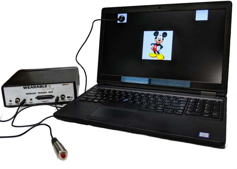
Our Wireless Trigger Hub simplifies the synchronization of DSI headsets with other devices. It features:
An additional benefit of the Trigger Hub design is that it allows synchronization across multiple data sources that are distributed across multiple systems, each of which running at its own clock rate. One such case commonly experienced in EEG experiments involves the synchronization of EEG and eye-tracking measurements, where the inevitable clock drift that arises between two systems during extended measurements creates difficulty in aligning data to events across the two systems.
The DSI-Flex can be customized so that an EEG sensor is replaced with a DSI auxiliary sensor. There are up to 7 locations on the DSI-Flex, enabling any configuration of the following sensors: EEG, ECG, EMG, EOG, GSR, RESP, & TEMP. The sensor data is collected and recorded in our data acquisition software, DSI-Streamer, where you can view the EEG and Aux sensors in real-time.
EEG Channels
Up to 7 Custom Sensor Locations
Reference / Ground
Common Mode Follower / Custom
Head Size Range
Custom Caps
Sampling Rate
300 Hz (600Hz upgrade available)
Bandwidth
0.003 – 150 Hz
A/D resolution
0.317 μV referred to input
Input Impedance (1Hz)
47 GΩ
CMRR
> 120 dB
Amplifier / Digitizer
16 bits / 7 channels
Wireless
Bluetooth
Wireless Range
10 m
Run-time
> 12 hours
Onboard Storage
~ 68 Hours (available option)
Data Acquisition
Real time, evoked potentials
Signal Quality Monitoring
Continuous impedance, Baseline offset, Noise (1-50 Hz)
Data Type
Raw and Filtered Data available
File Type
.CSV and .EDF
Data Output Streaming
TCP/IP socket, API (C Based), LSL
Cognitive State Classification
Brain Computer Interface
SSVEP BCI Algorithms; BCI2000; OpenViBE; PsychoPy; BCILab
Data Integration / Analysis
CAPTIV; Lab Streaming Layer; NeuroPype; BrainStorm; NeuroVIS
Neurofeedback
Applied Neuroscience NeuroGuide; Brainmaster Brain Avatar; EEGer
Neuromarketing
CAPTIV Neurolab
Presentation
Presentation; E-Prime
Mizrahi, Dor; Laufer, Ilan; Zuckerman, Inon
Modulation of Beta Power as a Function of Attachment Style and Feedback Valence Conference
International Conference on Brain Informatics, Springer 2023.
@conference{mizrahi2023modulation,

title = {Modulation of Beta Power as a Function of Attachment Style and Feedback Valence},

author = {Dor Mizrahi and Ilan Laufer and Inon Zuckerman},

url = {https://link.springer.com/chapter/10.1007/978-3-031-43075-6_2},

year  = {2023},

date = {2023-09-13},

urldate = {2023-01-01},

booktitle = {International Conference on Brain Informatics},

pages = {14–20},

organization = {Springer},

abstract = {Attachment theory is concerned with the basic level of social connection associated with approach and withdrawal mechanisms. Consistent patterns of attachment may be divided into two major categories: secure and insecure. As secure and insecure attachment style individuals vary in terms of their responses to affective stimuli and negatively valanced cues, the goal of this study was to examine whether there are differences in Beta power activation between secure and insecure individuals to feedback given while performing the arrow flanker task. An interaction emerged between Attachment style (secure or insecure) and Feedback type (success or failure) has shown differences in Beta power as a function of both independent factors. These results corroborate previous findings indicating that secure and insecure individuals differently process affective stimuli.},

keywords = {},

pubstate = {published},

tppubtype = {conference}

}
Georgiadis, Kostas; Kalaganis, Fotis P; Oikonomou, Vangelis P; Nikolopoulos, Spiros; Laskaris, Nikos A; Kompatsiaris, Ioannis
Harneshing the Potential of EEG in Neuromarketing with Deep Learning and Riemannian Geometry Conference
International Conference on Brain Informatics, Springer 2023.
@conference{georgiadis2023harneshing,

title = {Harneshing the Potential of EEG in Neuromarketing with Deep Learning and Riemannian Geometry},

author = {Kostas Georgiadis and Fotis P Kalaganis and Vangelis P Oikonomou and Spiros Nikolopoulos and Nikos A Laskaris and Ioannis Kompatsiaris},

url = {https://link.springer.com/chapter/10.1007/978-3-031-43075-6_3},

year  = {2023},

date = {2023-09-13},

urldate = {2023-01-01},

booktitle = {International Conference on Brain Informatics},

pages = {21–32},

organization = {Springer},

abstract = {Neuromarketing exploits neuroimaging techniques to study consumers’ responses to various marketing aspects, with the goal of gaining a more thorough understanding of the decision-making process. The neuroimaging technology encountered the most in neuromarketing studies is Electroencephalography (EEG), mainly due to its non-invasiveness, low cost and portability. Opposed to typical neuromarketing practices, which rely on signal-power related features, we introduce an efficient decoding scheme that is based on the principles of Riemannian Geometry and realized by means of a suitable deep learning (DL) architecture (i.e., SPDNet). We take advantage of a recently released, multi-subject, neuromarketing dataset to train SPDNet under the close-to-real-life scenario of product selection from a supermarket leaflet and compare its performance against standard tools in EEG-based neuromarketing. The sample covariance is used as an estimator of the ‘quasi-instantaneous’, brain activation pattern and derived from the multichannel signal recorded while the subject is gazing at a given product. Pattern derivation is followed by proper re-alignment to reduce covariate shift (inter-subject variability) before SPDNet casts its binary decision (i.e., “Buy”-“NoBuy”). The proposed decoder is characterized by sufficient generalizability to derive valid predictions upon unseen brain signals. Overall, our experimental results provide clear evidence about the superiority of the DL-decoder relatively to both conventional neuromarketing and alternative Riemannian Geometry-based approaches, and further demonstrate how neuromarketing can benefit from recent advances in data-centric machine learning and the availability of relevant experimental datasets.},

keywords = {},

pubstate = {published},

tppubtype = {conference}

}
Vall-llossera, Mónica Torrecilla; Farrens, Andria; Gupta, Disha; Reinkensmeyer, David
Electroencephalogram analysis of finger proprioception in healthy subjects Conference
International Brain Research Organization 2023.
@conference{nokey,

title = {Electroencephalogram analysis of finger proprioception in healthy subjects},

author = {Mónica Torrecilla Vall-llossera and Andria Farrens and Disha Gupta and David Reinkensmeyer

},

url = {https://wearablesensing.com/wp-content/uploads/2024/07/Poster_IBRO__Electroencephalogram_analysis_of_finger_proprioception_in_healthy_subjects_DEF_2.pdf},

year  = {2023},

date = {2023-09-09},

urldate = {2023-09-09},

organization = {International Brain Research Organization},

abstract = {Proprioception enables us to perceive the position and movement of our body parts, which is essential for motor control and performance. In this study, we investigated the neural processing underlying proprioception using electroencephalogram (EEG) signals during Crisscross, a robotic proprioception task. Nine healthy adults performed the task (aged 22-34) with their right-hand during EEG acquisition. In the Crisscross task, a robot crossed the index and middle fingers in an alternating flexion/extension pattern with vision of the hand occluded. Participants performed Crisscross in two modes; the Non Button Pressing mode, (CC-NBP), where participants simply relaxed the fingers as the robot moved them, and the Button pressing mode (CC-BP), where participants additionally pressed a button when they perceived their fingers were overlapped. We analysed the event-related potential (ERP) of EEG data at the instance of movement onset and of button pressing, and compared responses when participants were actively engaged in making decisions based on proprioception (CC-BP) to when they were not (CC-NBP). In both conditions, we observed a positive ERP in the P3 channel that was significantly larger in the CC-PB condition (MWU, p<0.001), indicating this response is modulated by participants attending to proprioceptive information. In the CC-BP condition observed a contingent negative variation (CNV) in the Cz channel that was time-locked to movement onset and peaked in magnitude at button-press, suggesting that it is associated with the proprioceptively-driven decision of when to press the button. Finally, both responses were larger when the robot moved the fingers at high speed (36 deg/s) compared to slow speeds (16 deg/s, p = 0.03). These EEG features provide insight into proprioceptive processing in healthy individuals, and allow us to assess sensorimotor function at the neural level. Future work will study these responses in post-stroke individuals to better understand sensorimotor deficits.},

keywords = {},

pubstate = {published},

tppubtype = {conference}

}
Georgiadis, Kostas; Kalaganis, Fotis P; Riskos, Kyriakos; Matta, Eleftheria; Oikonomou, Vangelis P; Yfantidou, Ioanna; Chantziaras, Dimitris; Pantouvakis, Kyriakos; Nikolopoulos, Spiros; Laskaris, Nikos A; others,
NeuMa-the absolute Neuromarketing dataset en route to an holistic understanding of consumer behaviour Journal Article
In: Scientific Data, vol. 10, no. 1, pp. 508, 2023.
@article{georgiadis2023neuma,

title = {NeuMa-the absolute Neuromarketing dataset en route to an holistic understanding of consumer behaviour},

author = {Kostas Georgiadis and Fotis P Kalaganis and Kyriakos Riskos and Eleftheria Matta and Vangelis P Oikonomou and Ioanna Yfantidou and Dimitris Chantziaras and Kyriakos Pantouvakis and Spiros Nikolopoulos and Nikos A Laskaris and others},

doi = {https://doi.org/10.1038/s41597-023-02392-9},

year  = {2023},

date = {2023-08-03},

urldate = {2023-01-01},

journal = {Scientific Data},

volume = {10},

number = {1},

pages = {508},

publisher = {Nature Publishing Group UK London},

abstract = {Neuromarketing is a continuously evolving field that utilises neuroimaging technologies to explore consumers’ behavioural responses to specific marketing-related stimulation, and furthermore introduces novel marketing tools that could complement the traditional ones like questionnaires. In this context, the present paper introduces a multimodal Neuromarketing dataset that encompasses the data from 42 individuals who participated in an advertising brochure-browsing scenario. In more detail, participants were exposed to a series of supermarket brochures (containing various products) and instructed to select the products they intended to buy. The data collected for each individual executing this protocol included: (i) encephalographic (EEG) recordings, (ii) eye tracking (ET) recordings, (iii) questionnaire responses (demographic, profiling and product related questions), and (iv) computer mouse data. NeuMa dataset has both dynamic and multimodal nature and, due to the narrow availability of open relevant datasets, provides new and unique opportunities for researchers in the field to attempt a more holistic approach to neuromarketing.},

keywords = {},

pubstate = {published},

tppubtype = {article}

}
Riek, Nathan T; Susam, Busra T; Hudac, Caitlin M; Conner, Caitlin M; Akcakaya, Murat; Yun, Jane; White, Susan W; Mazefsky, Carla A; Gable, Philip A
Feedback Related Negativity Amplitude is Greatest Following Deceptive Feedback in Autistic Adolescents Journal Article
In: Journal of Autism and Developmental Disorders, pp. 1–11, 2023.
@article{riek2023feedback,

title = {Feedback Related Negativity Amplitude is Greatest Following Deceptive Feedback in Autistic Adolescents},

author = {Nathan T Riek and Busra T Susam and Caitlin M Hudac and Caitlin M Conner and Murat Akcakaya and Jane Yun and Susan W White and Carla A Mazefsky and Philip A Gable},

url = {https://link.springer.com/article/10.1007/s10803-023-06038-y},

year  = {2023},

date = {2023-07-01},

urldate = {2023-01-01},

journal = {Journal of Autism and Developmental Disorders},

pages = {1–11},

publisher = {Springer},

abstract = {The purpose of this study is to investigate if feedback related negativity (FRN) can capture instantaneous elevated emotional reactivity in autistic adolescents. A measurement of elevated reactivity could allow clinicians to better support autistic individuals without the need for self-reporting or verbal conveyance. The study investigated reactivity in 46 autistic adolescents (ages 12–21 years) completing the Affective Posner Task which utilizes deceptive feedback to elicit distress presented as frustration. The FRN event-related potential (ERP) served as an instantaneous quantitative neural measurement of emotional reactivity. We compared deceptive and distressing feedback to both truthful but distressing feedback and truthful and non-distressing feedback using the FRN, response times in the successive trial, and Emotion Dysregulation Inventory (EDI) reactivity scores. Results revealed that FRN values were most negative to deceptive feedback as compared to truthful non-distressing feedback. Furthermore, distressing feedback led to faster response times in the successive trial on average. Lastly, participants with higher EDI reactivity scores had more negative FRN values for non-distressing truthful feedback compared to participants with lower reactivity scores. The FRN amplitude showed changes based on both frustration and reactivity. The findings of this investigation support using the FRN to better understand emotion regulation processes for autistic adolescents in future work. Furthermore, the change in FRN based on reactivity suggests the possible need to subgroup autistic adolescents based on reactivity and adjust interventions accordingly.},

keywords = {},

pubstate = {published},

tppubtype = {article}

}
Kim, Suhye; Kim, Jung-Hwan; Hyung, Wooseok; Shin, Suhkyung; Choi, Myoung Jin; Kim, Dong Hwan; Im, Chang-Hwan
Characteristic Behaviors of Elementary Students in a Low Attention State During Online Learning Identified Using Electroencephalography Journal Article
In: IEEE Transactions on Learning Technologies, 2023.
@article{kim2023characteristic,

title = {Characteristic Behaviors of Elementary Students in a Low Attention State During Online Learning Identified Using Electroencephalography},

author = {Suhye Kim and Jung-Hwan Kim and Wooseok Hyung and Suhkyung Shin and Myoung Jin Choi and Dong Hwan Kim and Chang-Hwan Im},

doi = {10.1109/TLT.2023.3289498},

year  = {2023},

date = {2023-06-29},

urldate = {2023-01-01},

journal = {IEEE Transactions on Learning Technologies},

publisher = {IEEE},

abstract = {With the widespread application of online education platforms, the necessity for identifying learner's mental states from webcam videos is increasing as it can be potentially applied to artificial intelligence-based automatic identification of learner's states. However, the behaviors that elementary school students frequently exhibit during online learning particularly when they are in a low attention state have rarely been investigated. This study employed electroencephalography (EEG) to continuously track changes in the learner's attention state during online learning. A new EEG index reflecting elementary students' attention level was developed using an EEG dataset acquired from 30 fourth graders during a computerized d2 test of attention. Characteristic behaviors of 24 elementary students in a low attention state were then identified from the webcam videos showing their upper bodies captured during 40-minute online lectures, with the proposed EEG index being used as a reference to determine their attention level at the time. Various characteristic behaviors were identified regarding participant's mouth, head, arms, and torso. For example, opening mouth or leaning back was observed more frequently in a low attention state than in a high attention state. It is expected that the characteristic behaviors reflecting learner's low attention state would be utilized as a useful reference in developing more interactive and effective online education systems.},

keywords = {},

pubstate = {published},

tppubtype = {article}

}
Cho, Ji Young; Wang, Ze-Yu; Hong, Yi-Kyung
An EEG Study on the Interior Environmental Design Elements in Promoting Healing Journal Article
In: Journal of the Korean Housing Association, vol. 34, no. 3, pp. 067–079, 2023.
@article{cho2023eeg,

title = {An EEG Study on the Interior Environmental Design Elements in Promoting Healing},

author = {Ji Young Cho and Ze-Yu Wang and Yi-Kyung Hong},

url = {https://journal.khousing.or.kr/articles/xml/XeZw/},

doi = {https://doi.org/10.6107/JKHA.2023.34.3.067},

year  = {2023},

date = {2023-06-25},

urldate = {2023-01-01},

journal = {Journal of the Korean Housing Association},

volume = {34},

number = {3},

pages = {067–079},

publisher = {The Korean Housing Association},

abstract = {This study aimed to identify the attributes of environmental design elements that promote healing. Eighteen college students participated in an EEG study, and their Ratio of Alpha to Beta (RAB) was examined in relation to eight interior design stimuli consisting of different conditions of illuminance (high/low), color (green/white), and biophilia ( natural/artificial). The EEGs of the 19 channels were measured using a dry-type EEG device (DSI-24). Participants also completed a survey questionnaire, choosing the best stimuli for healing environments and for wanting to stay. The EEG data were analyzed using a Wilcoxon signed-rank test to identify statistical differences in responses to different stimuli. The main results were as follows: (1) RAB was most frequently observed in the left temporal and parietal lobes; (2) when comparing pre- and post-stimulation, significant differences were observed between background EEG and six stimuli (S1, S3, S5, S6, S7, and S8); (3) RAB tended to be high with low illuminance, white walls, and artificial biophilia; and (4) the psychological and EEG responses did not match; stimuli with high illuminance and natural biophilia were selected the most for healing and wanting to stay environments. The results indicate the necessity for studying environmental responses via both physiological and psychological aspects as they compensate for each other. and artificial biophilia; and (4) the psychological and EEG responses did not match; stimuli with high illuminance and natural biophilia were selected the most for healing and wanting to stay environments. The results indicate the necessity for studying environmental responses via both physiological and psychological aspects as they compensate for each other. and artificial biophilia; and (4) the psychological and EEG responses did not match; stimuli with high illuminance and natural biophilia were selected the most for healing and wanting to stay environments. The results indicate the necessity for studying environmental responses via both physiological and psychological aspects as they compensate for each other.},

keywords = {},

pubstate = {published},

tppubtype = {article}

}
Fan, Tengwen; Zhang, Liming; Liu, Jianyi; Niu, Yanbin; Hong, Tian; Zhang, Wenfang; Shu, Hua; Zhao, Jingjing
Phonemic mismatch negativity mediates the association between phoneme awareness and character reading ability in young Chinese children Journal Article
In: Neuropsychologia, pp. 108624, 2023.
@article{fan2023phonemic,

title = {Phonemic mismatch negativity mediates the association between phoneme awareness and character reading ability in young Chinese children},

author = {Tengwen Fan and Liming Zhang and Jianyi Liu and Yanbin Niu and Tian Hong and Wenfang Zhang and Hua Shu and Jingjing Zhao},

doi = {https://doi.org/10.1016/j.neuropsychologia.2023.108624},

year  = {2023},

date = {2023-06-15},

urldate = {2023-01-01},

journal = {Neuropsychologia},

pages = {108624},

publisher = {Elsevier},

abstract = {Poor phonological awareness is associated with greater risk for reading disability. The underlying neural mechanism of such association may lie in the brain processing of phonological information. Lower amplitude of auditory mismatch negativity (MMN) has been associated with poor phonological awareness and with the presence of reading disability. The current study recorded auditory MMN to phoneme and lexical tone contrast with odd-ball paradigm and examined whether auditory MMN mediated the associations between phonological awareness and character reading ability through a three-year longitudinal study in 78 native Mandarin-speaking kindergarten children. Hierarchical linear regression and mediation analysis showed that the effect of phoneme awareness on the character reading ability was mediated by the phonemic MMN in young Chinese children. Findings underscore the key role of phonemic MMN for the underlying neurodevelopmental mechanism linking phoneme awareness and reading ability.},

keywords = {},

pubstate = {published},

tppubtype = {article}

}
Yu, Heeseung; Han, Eunkyoung
People see what they want to see: an EEG study Journal Article
In: Cognitive Neurodynamics, pp. 1–15, 2023.
@article{yu2023people,

title = {People see what they want to see: an EEG study},

author = {Heeseung Yu and Eunkyoung Han},

url = {https://link.springer.com/article/10.1007/s11571-023-09982-8},

year  = {2023},

date = {2023-06-13},

urldate = {2023-01-01},

journal = {Cognitive Neurodynamics},

pages = {1–15},

publisher = {Springer},

abstract = {This study explored selective exposure and confirmation bias in the choices participants made about which political videos to watch, and whether their political positions changed after they watched videos that either agreed with or opposed their positions on two controversial issues in South Korea: North Korea policy and social welfare policy. The participants completed questionnaires before and after they watched the videos, were asked to select thumbnails of videos before they watched any, and had their brain wave activity measured through electroencephalogram (EEG) as they watched both types of videos. The participants demonstrated selective exposure as they primarily selected video thumbnails with content that matched their political orientations, and they demonstrated confirmation bias as their questionnaire responses after they watched the videos indicated that their positions had hardened. There were also statistically significant differences in alpha, beta, sensory motor rhythm, low beta, mid beta, and fast alpha activity depending on the political orientation consistency between the participants and the videos. Future studies could expand this line of research beyond college students and beyond Asia, and longitudinal work could also be conducted to determine if the obtained patterns remain constant over time.},

keywords = {},

pubstate = {published},

tppubtype = {article}

}
Ferrisi, Leonardo M
Optimizing an assistive Brain Computer Interface that uses Auditory Attention as Input Masters Thesis
2023.
@mastersthesis{ferrisi2023optimizing,

title = {Optimizing an assistive Brain Computer Interface that uses Auditory Attention as Input},

author = {Leonardo M Ferrisi},

url = {https://digitalworks.union.edu/cgi/viewcontent.cgi?article=3754&context=theses},

year  = {2023},

date = {2023-06-01},

urldate = {2023-01-01},

abstract = {Brain Computer Interfaces (BCIs) allow individuals to operate technology using (typically consciously controllable) aspects of their brain activity. Auditory BCIs utilize principles of Auditory Event Related

Potentials or Auditory Evoked Potentials as a reproducible controllable features that individuals can use to operate a BCI. These Auditory BCIs in their most basic format can allow users to answer yes or no questions by listening to either one auditory stimuli or the other. Current accuracy in intended response detection for these kinds of BCIs can be as good as mean accuracy of 77 % [5]. BCI research tends to optimize the computer side of the system however the ease of use for the human operating the system is an important point of consideration as well. This research project aimed to determine what factors make a human operator able to achieve the highest accuracy using a given previously successfully demonstrated classifier. This research project primarily sought to answer the questions; to what degree people can improve their accuracy in operating an Auditory BCI and what factors of the stimulus used can be altered to achieve this. The results of this project, obtained through the data collected from six individuals, found that slower stimuli speeds for eliciting Auditory Event Related Potentials were significantly better at achieving higher prediction accuracies compared to faster stimulus speeds. The amount of time spent using the system appeared to result in diminishing returns in accuracy regardless of condition however not before an initial spike in greater classifier prediction accuracy for the second condition run on each subject. Although further research is needed to gain more conclusive evidence for or against the hypothesis, the results of this research may be able suggest that individuals can improve their performance using Auditory BCIs with practice at optimal parameters albeit within a given time frame before experiencing diminishing returns. These findings would stand to provide benefit both to continued research in making optimal non-invasive alternative communication technologies as well as making progress in finding the potential ceiling in accuracy that an Auditory BCI can have in interpreting brain activity for the intended action of the user},

keywords = {},

pubstate = {published},

tppubtype = {mastersthesis}

}
Please fill out the form and provide a brief description of your application so we can help match you with products that will meet your specific needs.
Please fill out the form and provide a brief description of your application so we can help match you with products that will meet your specific needs.