Wearable Sensing’s wireless DSI-Flex is the leading dry electrode EEG system in terms of signal quality and comfort. The DSI-Flex takes on average less than 5 minutes to set up, making it the ideal solution for scientists in need of a simple, easy to use, EEG system. Our patented sensor technology not only delivers uncompromised signal quality but also enables our system to be virtually immune against motion and electrical artifacts.
The DSI-Flex has dry sensors on flexible cables, enabling scientists to place the electrodes in varying configurations on the head. These flexible sensors are designed to be screwed into custom caps, so that scientists can order 1 DSI-Flex, and multiple caps, allowing for rapid application of multiple electrode configurations. Every sensor on the DSI-Flex can be customized as either ExG, GSR, TEMP, and REP. It also has a 4-bit trigger input to synchronize with other devices such as Eye-Tracking, Motion (IMU), and more.
Used around the world by leaders in Research, & Brain-Computer Interfaces
With over 90% correlation to research-grade wet EEG systems, the dry sensor interface (DSI) offers unparalleled quality and performance
Multiple adjustment points and a foam pad lined interior enable the system to be worn for up to 8 hours on any head shape or size
All DSI systems include free, unlimited licenses of DSI-Streamer, our data acquisition software which can record raw data, in .csv and .edf file formats
Faraday cage's, spring-loaded electrodes, and our patented common-mode follower technology, provides near immunity against electrical and motion artifacts
Using 70% isopropyl alcohol and a cleaning brush, the DSI-24 only takes a minute to clean, 3 minutes to dry, and can be up and running on the next subject in minutes
All DSI systems include our free C based .dll API, which enables users to pull the raw data directly from the headset, for custom software on Windows, Mac OS, Linux, and ARM
The DSI-Flex was designed for ultra-rapid setup, taking on average less than 5 minutes to don, and works on any type of hair, including long hair, thick hair, afros, and more
DSI headsets have active sensors, amplifiers, digitizers, batteries, onboard storage, and wireless transmission, making them complete, mobile, wearable EEG systems
DSI systems exclusively work with QStates, a machine learning algorithm for cognitive classification on states such as mental workload, engagement, and fatigue
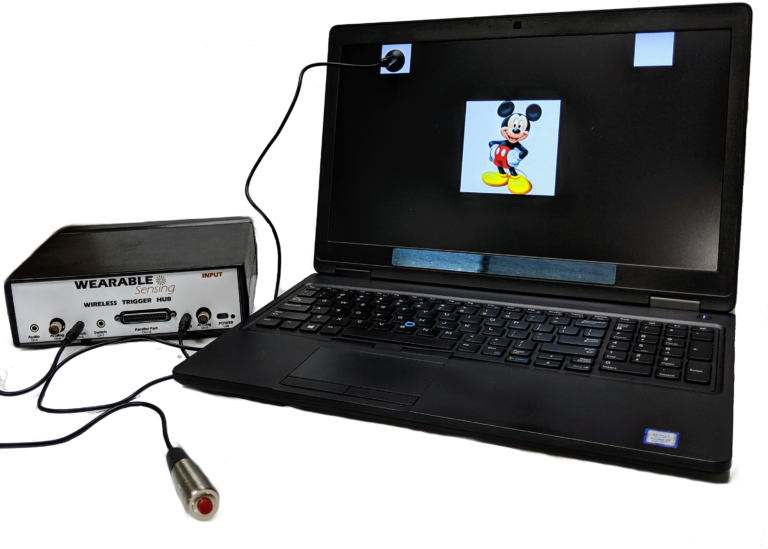
Our Wireless Trigger Hub simplifies the synchronization of DSI headsets with other devices. It features:
An additional benefit of the Trigger Hub design is that it allows synchronization across multiple data sources that are distributed across multiple systems, each of which running at its own clock rate. One such case commonly experienced in EEG experiments involves the synchronization of EEG and eye-tracking measurements, where the inevitable clock drift that arises between two systems during extended measurements creates difficulty in aligning data to events across the two systems.
The DSI-Flex can be customized so that an EEG sensor is replaced with a DSI auxiliary sensor. There are up to 7 locations on the DSI-Flex, enabling any configuration of the following sensors: EEG, ECG, EMG, EOG, GSR, RESP, & TEMP. The sensor data is collected and recorded in our data acquisition software, DSI-Streamer, where you can view the EEG and Aux sensors in real-time.
EEG Channels
Up to 7 Custom Sensor Locations
Reference / Ground
Common Mode Follower / Custom
Head Size Range
Custom Caps
Sampling Rate
300 Hz (600Hz upgrade available)
Bandwidth
0.003 – 150 Hz
A/D resolution
0.317 μV referred to input
Input Impedance (1Hz)
47 GΩ
CMRR
> 120 dB
Amplifier / Digitizer
16 bits / 7 channels
Wireless
Bluetooth
Wireless Range
10 m
Run-time
> 12 hours
Onboard Storage
~ 68 Hours (available option)
Data Acquisition
Real time, evoked potentials
Signal Quality Monitoring
Continuous impedance, Baseline offset, Noise (1-50 Hz)
Data Type
Raw and Filtered Data available
File Type
.CSV and .EDF
Data Output Streaming
TCP/IP socket, API (C Based), LSL
Cognitive State Classification
Brain Computer Interface
SSVEP BCI Algorithms; BCI2000; OpenViBE; PsychoPy; BCILab
Data Integration / Analysis
CAPTIV; Lab Streaming Layer; NeuroPype; BrainStorm; NeuroVIS
Neurofeedback
Applied Neuroscience NeuroGuide; Brainmaster Brain Avatar; EEGer
Neuromarketing
CAPTIV Neurolab
Presentation
Presentation; E-Prime
Matavalam, Chandu Priya; Gangadharan, Sagila; Vinod, AP
Consumer Trait Prediction from EEG Using a Novel SpectralTraitNet for Neuromarketing Application Conference
2026 IEEE International Conference on Human-Machine Systems (ICHMS), IEEE 2026.
@conference{matavalam2026consumer,

title = {Consumer Trait Prediction from EEG Using a Novel SpectralTraitNet for Neuromarketing Application},

author = {Chandu Priya Matavalam and Sagila Gangadharan and AP Vinod},

doi = {https://doi.org/10.1109/ICHMS69701.2026.11602183},

year  = {2026},

date = {2026-07-15},

urldate = {2026-01-01},

booktitle = {2026 IEEE International Conference on Human-Machine Systems (ICHMS)},

pages = {416–421},

organization = {IEEE},

abstract = {Decoding stable consumer traits from Electroencephalography (EEG) remains a largely unexplored frontier in neuromarketing, which has traditionally focused on short-term preference classification (e.g., “Like” vs. “Dislike”). Moving beyond transient reactions to identifying long-term profiles, such as the “Bargain Hunter”- a consumer predominantly motivated by finding the best deals and discounts to make purchase decisions, enables marketers to target promotions more effectively to drive conversions. However, consumer trait prediction is challenging due to the subtle nature of trait-based neural signatures, intra-subject label homogeneity, and significant inter-subject variability. Unlike conscious preference tasks, consumer traits function as subconscious priors, resulting in a high risk of the model overfitting to individual identities rather than generalized traits. In this study, we propose a subject-independent deep learning framework to classify “Bargain Hunter” vs. “Non-Bargain Hunter” traits using EEG signals recorded during product browsing. A hybrid lightweight Convolutional Neural Network derived by combining EEGNet and MobileNet, called SpectralTraitNet, which uses depthwise separable convolutions and attention mechanism, is proposed to extract discriminative spatial-spectral features. To enhance cross-subject generalization, Euclidean Alignment is employed to align subject-specific covariance matrices into a common manifold, effectively addressing domain shifts. The framework is evaluated on the publicly available NeuMa dataset (N=42), and a subject-level classification accuracy of 6 4. 2 9±1 8 % is achieved using rigorous 6 -fold stratified cross-validation. These results demonstrate that manifold alignment and data augmentation are critical for extracting generalized features across unseen subjects, offering a pathway towards more personalized, neurologically informed pricing and marketing strategies.},

keywords = {},

pubstate = {published},

tppubtype = {conference}

}
Kim, H; Jun, SC; Nam, CS
A reproducible EEG hyperscanning dataset for triadic social decision-making during an iterated 3-player Prisoner’s Dilemma Journal Article
In: Data in Brief, pp. 113064, 2026.
@article{kim2026reproducible,

title = {A reproducible EEG hyperscanning dataset for triadic social decision-making during an iterated 3-player Prisoner’s Dilemma},

author = {H Kim and SC Jun and CS Nam},

doi = {https://doi.org/10.1016/j.dib.2026.113064},

year  = {2026},

date = {2026-07-10},

urldate = {2026-01-01},

journal = {Data in Brief},

pages = {113064},

publisher = {Elsevier},

abstract = {This data article describes an open EEG hyperscanning dataset acquired during an iterated 3-player Prisoner’s Dilemma (PD) task, designed to support reproducible research on triadic social decision-making. EEG was recorded simultaneously from three participants per group (11 groups; 33 subjects) using synchronized acquisition, with decision-locked and feedback-locked epochs provided for 40 task trials per subject and three 60-s resting-state runs per group. The released MATLAB files contain stacked 57-channel EEG arrays (19 channels × 3 subjects) sampled at 300 Hz, with explicit epoch definitions for decision (−1000 to 4000 ms) and feedback (−1000 to 2000 ms) periods, as well as trial-wise behavioral choice labels (1=cooperate, 2=defect) enabling reconstruction of dyad- and triad-level outcomes. Questionnaire metadata (personal information and pre/mid/post state measures) Yare also provided as supplementary spreadsheets. To facilitate reuse and benchmarking, we release Python code that reproduces the preprocessing pipeline (average reference, FIR filtering, ICA + ICLabel for task EEG), ERP summaries, and inter-brain synchrony measures (PLV and coherence) with window-matched resting baselines and cluster-permutation statistics. The dataset is intended for method development and benchmarking in ERP analysis, inter-brain synchrony estimation, and modeling of dynamic group interaction states in triadic games.},

keywords = {},

pubstate = {published},

tppubtype = {article}

}
Debnath, Shubham; Fylaktou, Fylaktis; Gurfein, Blake T; Zanos, Theodoros P
Autonomic and neural responses to varying transcutaneous cervical electrical stimulation parameters Journal Article
In: Bioelectronic Medicine, vol. 12, no. 1, pp. 16, 2026.
@article{debnath2026autonomic,

title = {Autonomic and neural responses to varying transcutaneous cervical electrical stimulation parameters},

author = {Shubham Debnath and Fylaktis Fylaktou and Blake T Gurfein and Theodoros P Zanos},

doi = {https://doi.org/10.1186/s42234-026-00210-2},

year  = {2026},

date = {2026-07-06},

urldate = {2026-07-06},

journal = {Bioelectronic Medicine},

volume = {12},

number = {1},

pages = {16},

publisher = {Springer},

abstract = {Background

Transcutaneous cervical electrical stimulation (TCES) offers a noninvasive approach to modulate the autonomic nervous system (ANS), but optimal stimulation parameters remain undefined. This pilot study aimed to identify optimal TCES parameters by evaluating autonomic and neural responses across varying frequencies, current intensities, electrode montages, and durations, using heart rate variability (HRV) and electroencephalography (EEG) alpha-band power as biomarkers of parasympathetic activity.


Methods

Twenty healthy adults completed four testing sessions, each examining one stimulation parameter. Autonomic data were collected including electrocardiography, non-invasive blood pressure, pupillometry, photoplethysmography, and dry-electrode EEG. Four frequencies (10, 25, 40, 150 Hz), three current intensities (sub-sensation threshold, sensation threshold, supra-sensation threshold), three electrode montages (bilateral, left-only, right-only), and two durations (4, 20 min) were tested. Root mean square of successive differences (RMSSD) and global EEG alpha-band power were primary outcomes. Parameters were sequentially optimized across visits based on individual RMSSD responses.


Results

No single frequency produced a significantly higher RMSSD or alpha-band power response. However, each participant exhibited a personalized preferred frequency yielding a mean 41% RMSSD increase in visit 1. This individualized frequency was selected for further visits varying current intensity and electrode montage. Supra-sensation threshold intensity was most effective, with 60% of participants responding strongest at this level. Left-sided stimulation resulted in a decrease in both RMSSD and alpha-band power, while right-sided and bilateral montages resulted in similar increases for these biomarkers. Due to decreasing cardiac vagal response in successive sessions, the preferred frequency was reevaluated before testing duration. The mean RMSSD response increased 54% upon recalibration in visit 4, though the preferred frequency shifted in 75% of participants. Autonomic vitals did not significantly modulate more with longer stimulation duration; pulse rate variability during 20-min stimulation revealed oscillatory autonomic dynamics with peak parasympathetic responses emerging around 4 min.


Conclusions

TCES can modulate cardiac vagal and cortical responses as measured by RMSSD and EEG alpha-band power, respectively, and a personalized, biomarker-guided approach to TCES parameter optimization is essential for future clinical applications targeting autonomic dysfunction.},

keywords = {},

pubstate = {published},

tppubtype = {article}

}
Xu, Chi; Li, Xiang; Shehata, Allam; Aljazaerly, Mohamad Ammar Alsherfawi; Yagi, Yasushi
OU-MB: The OU Multimodal Biometric Database and Its Performance Evaluation Journal Article
In: IEEE Transactions on Biometrics, Behavior, and Identity Science, 2026.
@article{xu2026mb,

title = {OU-MB: The OU Multimodal Biometric Database and Its Performance Evaluation},

author = {Chi Xu and Xiang Li and Allam Shehata and Mohamad Ammar Alsherfawi Aljazaerly and Yasushi Yagi},

doi = {https://doi.org/10.1109/TBIOM.2026.3710514},

year  = {2026},

date = {2026-07-06},

urldate = {2026-01-01},

journal = {IEEE Transactions on Biometrics, Behavior, and Identity Science},

publisher = {IEEE},

abstract = {In this paper, we describe a new multimodal biometric database named “the University of Osaka Multimodal Biometric Database (OU-MB)”. This database consists of 1,099 subjects and eleven biometric modalities, which, to the best of our knowledge, is the largest number of modalities among existing multimodal databases. Specifically, for each subject, we collected his/her irises, palm veins, 2D face images, signature images, gait videos, and voice data, which are typically included in existing multimodal databases. Additionally, some modalities not commonly considered in previous datasets are also included, that is, full-body images, online signature time series data, brain signals, inertial data (e.g., acceleration), and health data (e.g., heartbeat). We provide comprehensive baseline evaluations across all eleven biometric modalities included in OU-MB and further investigate multimodal recognition through representative score-level and feature-level fusion experiments. We believe this database can facilitate future research on person authentication using unimodal, multimodal, and even cross-modal approaches, as well as research on brain signal and health status analysis.

},

keywords = {},

pubstate = {published},

tppubtype = {article}

}
Zhou, Qinyu; Ng, Kam KH
Establishing Human Factors-Driven No-Fly Zone for Manned eVTOLs in Urban Air Mobility Conference
International Conference on Human-Computer Interaction, Springer 2026.
@conference{zhou2026establishing,

title = {Establishing Human Factors-Driven No-Fly Zone for Manned eVTOLs in Urban Air Mobility},

author = {Qinyu Zhou and Kam KH Ng},

doi = {https://doi.org/10.1007/978-3-032-29456-2_22},

year  = {2026},

date = {2026-06-22},

urldate = {2026-06-22},

booktitle = {International Conference on Human-Computer Interaction},

pages = {305–317},

organization = {Springer},

abstract = {Spatial envelop design of no-fly zone (NFZ) in urban air mobility (UAM) considers various factors and constraints. This study proposed a new NFZ boundary design method taking human factors into consideration. An obstacle avoiding experiment was conducted under 4 levels of distances including 104, 78, 52 and 26 m. Psychological, physiological and behavioral data were all recorded from the experiment to reflect stress levels of the participants under different building avoiding distances. Then a multi-criteria decision making (MCDM) framework was proposed to calculate the composite stress indicator (CSI) through the fusion of the three data sources. The two-order differences of CSI values were calculated to find out the turning point of stress. The result showed that CSIs from the 4 levels of distances were computed as 0.066, 0.100, 0.324 and 1.000. The 52-m building avoiding distance witnessed the abrupt change of CSI, indicating that 52-m separation can be regarded as a safety margin for establishing stress-driven no-fly zone. This contributes to the safe separation design in urban airspace.},

keywords = {},

pubstate = {published},

tppubtype = {conference}

}
Jeong, Eunju; Hong, You Jeong; Shin, Jiyeon; Kim, Jong Su; Yoo, Moon A; Kim, Sung-Phil
Psychological Empowerment on the Streets: Designing and Validating Multisensory Experiences in Simulated Autonomous Driving Journal Article
In: Annals of the New York Academy of Sciences, vol. 1560, no. 1, pp. e70305, 2026.
@article{jeong2026psychological,

title = {Psychological Empowerment on the Streets: Designing and Validating Multisensory Experiences in Simulated Autonomous Driving},

author = {Eunju Jeong and You Jeong Hong and Jiyeon Shin and Jong Su Kim and Moon A Yoo and Sung-Phil Kim},

doi = {https://doi.org/10.1111/nyas.70305},

year  = {2026},

date = {2026-06-16},

urldate = {2026-01-01},

journal = {Annals of the New York Academy of Sciences},

volume = {1560},

number = {1},

pages = {e70305},

publisher = {Wiley Online Library},

abstract = {Driving is evolving from a transportation task into a rich, multisensory experience in automated vehicles. This study developed and evaluated three multisensory solutions combining music with synchronized vibrotactile stimulation for autonomous driving contexts: safe (city driving), engagement (highway cruising), and entertainment (highway entry). Eighteen healthy adult drivers experienced three context–solution pairs presented in music only (M) and music with vibration (MV) modalities with simulated autonomous driving scenarios. Participants’ responses were measured using self-assessment manikin (SAM) ratings and electroencephalography (EEG). A significant main effect of modality showed that MV led to greater pleasure than M (EEG: p < 0.05; SAM: p < 0.05), with arousal showing a similar pattern (EEG: p < 0.05; SAM: p = 0.099). Behavioral data showed different emotional profiles across the three context−solution pairs (p < 0.001 for pleasure, arousal, and dominance), whereas the EEG contrast, which subtracted the video-only condition, showed no significant pair effect. These findings demonstrate that vibrotactile enhancement provides consistent emotional benefits across diverse driving contexts. Because each musical solution was paired with a unique driving scenario, these differences cannot be attributed solely to the music intervention. Future optimization of music and vibrotactile parameters may further enhance the autonomous driving experience.},

keywords = {},

pubstate = {published},

tppubtype = {article}

}
Susam, Busra T; Riek, Nathan T; Gall, Richard T; Conner, Caitlin M; White, Susan W; Mazefsky, Carla A; Akcakaya, Murat
Neural effects of meditation following a randomized controlled trial of the Emotion Awareness and Skills Enhancement (EASE) Journal Article
In: IEEE Transactions on Neural Systems and Rehabilitation Engineering, 2026.
@article{susam2026neural,

title = {Neural effects of meditation following a randomized controlled trial of the Emotion Awareness and Skills Enhancement (EASE)},

author = {Busra T Susam and Nathan T Riek and Richard T Gall and Caitlin M Conner and Susan W White and Carla A Mazefsky and Murat Akcakaya},

doi = {https://doi.org/10.1109/TNSRE.2026.3698744},

year  = {2026},

date = {2026-06-01},

urldate = {2026-01-01},

journal = {IEEE Transactions on Neural Systems and Rehabilitation Engineering},

publisher = {IEEE},

abstract = {Mindfulness has promise for enhancing emotion regulation in autistic individuals. Electroencephalography (EEG) emerges as an ideal, objective measure of neural responses before and after mindfulness practice. In a study with 39 autistic individuals, EEG analysis assessed the impact of mindfulness in the participants in a randomized controlled clinical trial of the Emotion Awareness and Skills Enhancement (EASE) program compared to a control group. Participants completed an affective Posner Task while wearing an EEG headset during two visits. In between visits, each participant either received EASE or active control therapy. Using random forest classifiers over the Post-Visit EEG features baseline corrected by Pre-Visit, the study achieved 88.48% to 91.86% accuracy in distinguishing EASE and Control groups under distress, non-distress, and neutral conditions. Linear mixed-effects models applied across the full EEG feature set revealed significant Visit × Feedback × Group interactions in central theta and midline and occipital beta band power, with post-hoc analyses indicating these effects were primarily driven by differential neural responses to rewarding versus unfavorable feedback. The EASE group demonstrated distinct pre-to-post changes in these features relative to the Control group, suggesting intervention-related modulation of neural systems supporting adaptive responses to emotionally meaningful feedback. These findings underscore mindfulness' positive influence on emotion regulation potentially showing its effects as neural oscillations matching the results from existing literature. Subgroup analysis based on Clinical Global Impressions threshold scores identified responders and non-responders with LME analyses confirming greater intervention-related EEG changes in EASE responders compared to Control responders.Our findings provide valuable insights into potential benefits of mindfulness-based interventions for autistic people, highlighting the neurophysiological effects of such programs.

},

keywords = {},

pubstate = {published},

tppubtype = {article}

}
Fincham, Jon M; Betts, Shawn; Anderson, John R
Combining EEG signals from the 2 members of a team to improve event identification Journal Article
In: NeuroImage: Reports, vol. 6, no. 2, pp. 100356, 2026.
@article{fincham2026combining,

title = {Combining EEG signals from the 2 members of a team to improve event identification},

author = {Jon M Fincham and Shawn Betts and John R Anderson},

doi = {https://doi.org/10.1016/j.ynirp.2026.100356},

year  = {2026},

date = {2026-05-20},

urldate = {2026-05-20},

journal = {NeuroImage: Reports},

volume = {6},

number = {2},

pages = {100356},

publisher = {Elsevier},

abstract = {We examined the potential of combining EEG signals from multiple individuals to identify critical events in a team task. In this study two subjects played a video game in which they had complementary roles, one player serving as a Bait to distract 5 enemy fortress and the other serving as a Shooter to destroy the fortress. Twenty-one pairs of subjects were analyzed. Critical events, destruction of the fortress and deaths of each player, evoked distinguishable P300-like responses from both players. Fortress kills could be best identified by combining the two EEG signals, while deaths could be best identified by focusing on the response of the player who died. Hidden semi-Markov models (HSMMs) achieved good identification of the events by combining information about the temporal distribution of these critical events with the conditional probability of the EEG activity. These findings indicate that we can track and improve by adaptively merging or selecting the signals from different team members.

},

keywords = {},

pubstate = {published},

tppubtype = {article}

}
Gravunder, Andrew; Studnicki, Amanda; Kline, Julia; Behboodi, Ahad; Bulea, Thomas C; Damiano, Diane L
In: Bioengineering, vol. 13, no. 5, pp. 561, 2026.
@article{gravunder2026novel,

title = {Novel Time-Series Forecasting Method to Enhance Accuracy of Real-Time EEG Detection for BCI-Based Neurofeedback Motor Training in Individuals with Cerebral Palsy and Other Neurological Disorders},

author = {Andrew Gravunder and Amanda Studnicki and Julia Kline and Ahad Behboodi and Thomas C Bulea and Diane L Damiano},

doi = {https://doi.org/10.3390/bioengineering13050561},

year  = {2026},

date = {2026-05-15},

urldate = {2026-01-01},

journal = {Bioengineering},

volume = {13},

number = {5},

pages = {561},

publisher = {MDPI},

abstract = {Real-time detection of motor intent using electroencephalography (EEG) with high accuracy remains a technical challenge for neurorehabilitation. Brain–computer interface-based neurofeedback training (BCI-NFT) paradigms need to detect pre-movement EEG to activate robotics or electrical stimulation nearly simultaneously with movement to promote neuroplasticity. We present a novel detection method commonly used in time-series forecasting (e.g., stock market trends), identifying crosses in fast (short) and slow (long) moving average windows to identify negative deflections in slow movement-related cortical potentials (MRCPs) or event-related desynchronization (ERD) within −400–+100 ms of movement onset. We recorded EEG data from the Cz electrode during our cued ankle dorsiflexion BCI-NFT paradigm in four adult participants, two neurotypical and two with cerebral palsy. Simulated real-time offline analyses demonstrated an 85.9% mean true positive rate and 14.1% false positive rate of detecting motor intent at a mean −182 ms from movement onset. We further evaluated whether the detection indicated a MRCP and/or ERD, with MRCP detected in 70–80% of trials in three participants, but high ERD detection (87%) instead in the other. Preliminary results indicate that this approach offers a straightforward, accurate, and well-timed method for real-time EEG detection during neurofeedback training and as a control signal for brain–computer interfaces.

},

keywords = {},

pubstate = {published},

tppubtype = {article}

}
Wang, Jun; Wu, Yibo; Xu, Jihong; Xie, Jiatong; Li, Zanyang; Imtiaz, Muhammad
An Adaptive Dynamic Window Strategy for SSVEP Identification Based on FBCCA Conference
2026 9th International Conference on Advanced Algorithms and Control Engineering (ICAACE), IEEE 2026.
@conference{wang2026adaptive,

title = {An Adaptive Dynamic Window Strategy for SSVEP Identification Based on FBCCA},

author = {Jun Wang and Yibo Wu and Jihong Xu and Jiatong Xie and Zanyang Li and Muhammad Imtiaz},

doi = {https://doi.org/10.1109/ICAACE69793.2026.11509205},

year  = {2026},

date = {2026-05-13},

urldate = {2026-01-01},

booktitle = {2026 9th International Conference on Advanced Algorithms and Control Engineering (ICAACE)},

pages = {2527–2531},

organization = {IEEE},

abstract = {Standard Filter Bank Canonical Correlation Analysis (FBCCA) utilizes fixed time windows, which limits adaptability to individual Steady-State Visual Evoked Potential (SSVEP) variability and restricts performance. To address this, this paper proposes a Dynamic Window Strategy FBCCA (FBCCA-DS) algorithm that adaptively determines the optimal data length using a threshold-based stopping strategy. By optimizing the trade-off between detection speed and accuracy, the proposed method significantly outperforms fixed-window baselines. Experimental results demonstrate an average offline accuracy of 86.54% and an Information Transfer Rate (ITR) of 115.04 bits/min, with comparable online performance (88.15% accuracy, 114.75 bits/min ITR). These findings indicate that the dynamic strategy effectively enhances the robustness and efficiency of SSVEP-based Brain-Computer Interface (BCI) systems.},

keywords = {},

pubstate = {published},

tppubtype = {conference}

}