Wearable Sensing’s wireless DSI-7 is the leading dry electrode EEG system in terms of signal quality and comfort. The DSI-7 takes on average less than 1 minute to set up, making it the ideal solution for scientists in need of a simple, easy to use, EEG system. Our patented sensor technology not only delivers uncompromised signal quality but also enables our system to be virtually immune against motion and electrical artifacts. As a result, the DSI-7 can be utilized in virtual or augmented reality, while also allowing researchers to take their experiments out of the lab, and into the real world.
The DSI-7 has sensor locations covering the frontal, center, and posterior areas of the brain. There are 7 sensors whose locations can be customized upon order, and 2 earclip sensors. It also has a 4-bit trigger input to synchronize with other devices such as Eye-Tracking, Motion (IMU), and more.
Used around the world by leaders in Research, Neurofeedback, Neuromarketing, Brain-Computer Interfaces, & Neuroergonomics.
With over 90% correlation to research-grade wet EEG systems, the dry sensor interface (DSI) offers unparalleled quality and performance
Multiple adjustment points and a foam pad lined interior enable the system to be worn for up to 8 hours on any head shape or size
All DSI systems include free, unlimited licenses of DSI-Streamer, our data acquisition software which can record raw data, in .csv and .edf file formats
Faraday cage's, spring-loaded electrodes, and our patented common-mode follower technology, provides near immunity against electrical and motion artifacts
Using 70% isopropyl alcohol and a cleaning brush, the DSI-24 only takes a minute to clean, 3 minutes to dry, and can be up and running on the next subject in minutes
All DSI systems include our free C based .dll API, which enables users to pull the raw data directly from the headset, for custom software on Windows, Mac OS, Linux, and ARM
The DSI-7 was designed for ultra-rapid setup, taking on average less than 1 minute to don, and works on any type of hair, including long hair, thick hair, afros, and more
DSI headsets have active sensors, amplifiers, digitizers, batteries, onboard storage, and wireless transmission, making them complete, mobile, wearable EEG systems
DSI systems exclusively work with QStates, a machine learning algorithm for cognitive classification on states such as mental workload, engagement, and fatigue
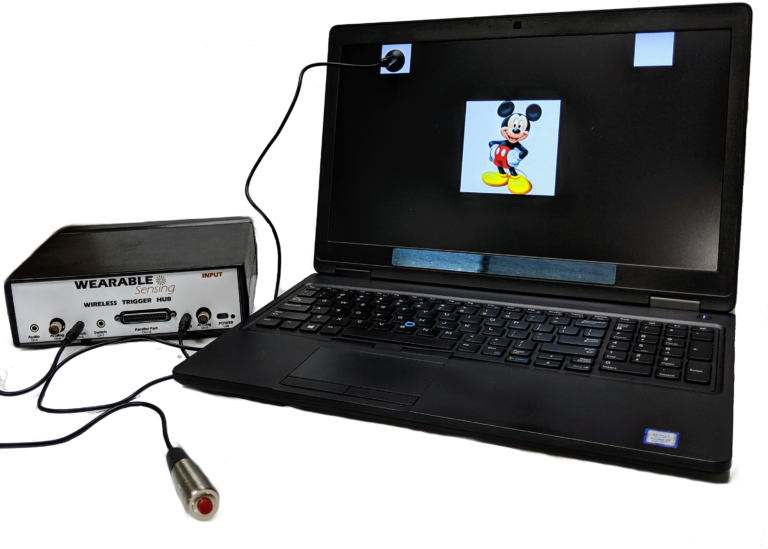
Our Wireless Trigger Hub simplifies the synchronization of DSI headsets with other devices. It features:
An additional benefit of the Trigger Hub design is that it allows synchronization across multiple data sources that are distributed across multiple systems, each of which running at its own clock rate. One such case commonly experienced in EEG experiments involves the synchronization of EEG and eye-tracking measurements, where the inevitable clock drift that arises between two systems during extended measurements creates difficulty in aligning data to events across the two systems.
The DSI-7 can be customized to have ECG, EMG, EOG, GSR, RESP, & TEMP. To do this, you can choose to either remove an EEG sensor in exchange for an auxiliary sensor, or you can modify the earclips to record ExG. The sensor data is collected and recorded in our data acquisition software, DSI-Streamer, where you can view the EEG and Aux sensors in real-time.
EEG Channels
Can be customized on demand by manufacturer
Reference / Ground
Common Mode Follower / Fz
Head Size Range
Adult Size: 52cm – 62cm circumference
Child Size: 48cm – 54cm circumference
Sampling Rate
300 Hz (600Hz upgrade available)
Bandwidth
0.003 – 150 Hz
A/D resolution
0.317 μV referred to input
Input Impedance (1Hz)
47 GΩ
CMRR
> 120 dB
Amplifier / Digitizer
16 bits / 7 channels
Wireless
Bluetooth
Wireless Range
10 m
Run-time
> 12 Hours
Onboard Storage
~ 68 Hours (available option)
Data Acquisition
Real time, evoked potentials
Signal Quality Monitoring
Continuous impedance, Baseline offset, Noise (1-50 Hz)
Data Type
Raw and Filtered Data available
File Type
.CSV and .EDF
Data Output Streaming
TCP/IP socket, API (C Based), LSL
Cognitive State Classification
Brain Computer Interface
SSVEP BCI Algorithms; BCI2000; OpenViBE; PsychoPy; BCILab
Data Integration / Analysis
CAPTIV; Lab Streaming Layer; NeuroPype; BrainStorm; NeuroVIS
Neurofeedback
Brainmaster Brain Avatar; EEGer
Neuromarketing
CAPTIV Neurolab
Presentation
Presentation; E-Prime
Dong, Xian; Wu, Yeyu; Tu, Zhijun; Cao, Bin; Li, Xianting; Yang, Zixu; Liu, Fei; Xing, Zheli
Influence of ambient temperature on personnel thermal comfort and working efficiency under isolation condition of underground engineering Journal Article
In: Energy and Buildings, pp. 112438, 2022.
@article{dong2022influence,

title = {Influence of ambient temperature on personnel thermal comfort and working efficiency under isolation condition of underground engineering},

author = {Xian Dong and Yeyu Wu and Zhijun Tu and Bin Cao and Xianting Li and Zixu Yang and Fei Liu and Zheli Xing},

doi = {https://doi.org/10.1016/j.enbuild.2022.112438},

year  = {2022},

date = {2022-08-29},

urldate = {2022-01-01},

journal = {Energy and Buildings},

pages = {112438},

publisher = {Elsevier},

abstract = {When attacked by weapons of mass destruction, underground engineering will operate under isolation condition, which results in the increase of temperature, humidity, and CO2 concentration. At present, there are few studies on personnel thermal comfort and working efficiency in underground engineering, especially under isolation condition. To improve the personnel thermal comfort and working efficiency under such condition, the influence of environmental temperature changes on human thermal comfort and working efficiency was investigated through a combination of subjective and objective methods. A subjective questionnaire and working efficiency test were conducted on the subjects in the artificial climate chamber, and synchronously monitored electrocardiography (ECG), electroencephalography (EEG), and other physiological parameters of the subjects were recorded, when isolation condition was achieved in the artificial climate chamber. The results show that: (1) the human neutral temperature is 24.2 °C, and thermal comfort zone is [23 °C, 25.5 °C] for isolation condition; (2) the high working efficiency area is [27.3 °C, 28.8 °C] for isolation condition; (3) the average of the TSV corresponding to the highest working efficiency point is 1.3 under isolation condition; (4) from the correlation analysis of working efficiency and personnel physiological indicators, personnel EEG index and task performance are significantly related, and the ECG index and task performance are not relevant for subjects performing brain work under isolation conditions.},

keywords = {},

pubstate = {published},

tppubtype = {article}

}
Humphries, Joseph B; Mattos, Daniela JS; Rutlin, Jerrel; Daniel, Andy GS; Rybczynski, Kathleen; Notestine, Theresa; Shimony, Joshua S; Burton, Harold; Carter, Alexandre; Leuthardt, Eric C
Motor Network Reorganization Induced in Chronic Stroke Patients with the Use of a Contralesionally-Controlled Brain Computer Interface Journal Article
In: Brain-Computer Interfaces, vol. 9, no. 3, pp. 179–192, 2022.
@article{humphries2022motor,

title = {Motor Network Reorganization Induced in Chronic Stroke Patients with the Use of a Contralesionally-Controlled Brain Computer Interface},

author = {Joseph B Humphries and Daniela JS Mattos and Jerrel Rutlin and Andy GS Daniel and Kathleen Rybczynski and Theresa Notestine and Joshua S Shimony and Harold Burton and Alexandre Carter and Eric C Leuthardt},

doi = {https://doi.org/10.1080/2326263X.2022.2057757},

year  = {2022},

date = {2022-07-01},

urldate = {2022-01-01},

journal = {Brain-Computer Interfaces},

volume = {9},

number = {3},

pages = {179--192},

publisher = {Taylor & Francis},

abstract = {Upper extremity weakness in chronic stroke remains a problem not fully addressed by current therapies. Brain–computer interfaces (BCIs) engaging the unaffected hemisphere are a promising therapy that are entering clinical application, but the mechanism underlying recovery is not well understood. We used resting state functional MRI to assess the impact a contralesionally driven EEG BCI therapy had on motor system functional organization. Patients used a therapeutic BCI for 12 weeks at home. We acquired resting-state fMRI scans and motor function data before and after the therapy period. Changes in functional connectivity (FC) strength between motor network regions of interest (ROIs) and the topographic extent of FC to specific ROIs were analyzed. Most patients achieved clinically significant improvement. Motor FC strength and topographic extent decreased following BCI therapy. Motor recovery correlated with reductions in motor FC strength across the entire motor network. These findings suggest BCI-mediated interventions may reverse pathologic strengthening of dysfunctional network interactions.},

keywords = {},

pubstate = {published},

tppubtype = {article}

}
Rustamov, Nabi; Humphries, Joseph; Carter, Alexandre; Leuthardt, Eric C
Theta-gamma coupling as a cortical biomarker of brain-computer interface mediated motor recovery in chronic stroke Journal Article
In: Brain Communications, vol. 4, iss. 3, 2022.
@article{rustamov2022thetab,

title = {Theta-gamma coupling as a cortical biomarker of brain-computer interface mediated motor recovery in chronic stroke},

author = {Nabi Rustamov and Joseph Humphries and Alexandre Carter and Eric C Leuthardt},

doi = {https://doi.org/10.1093/braincomms/fcac136},

year  = {2022},

date = {2022-05-25},

urldate = {2022-01-01},

journal = {Brain Communications},

volume = {4},

issue = {3},

abstract = {Chronic stroke patients with upper-limb motor disabilities are now beginning to see treatment options that were not previously available. To date, the two options recently approved by the United States Food and Drug Administration include vagus nerve stimulation and brain–computer interface therapy. While the mechanisms for vagus nerve stimulation have been well defined, the mechanisms underlying brain–computer interface-driven motor rehabilitation are largely unknown. Given that cross-frequency coupling has been associated with a wide variety of higher-order functions involved in learning and memory, we hypothesized this rhythm-specific mechanism would correlate with the functional improvements effected by a brain–computer interface. This study investigated whether the motor improvements in chronic stroke patients induced with a brain–computer interface therapy are associated with alterations in phase–amplitude coupling, a type of cross-frequency coupling. Seventeen chronic hemiparetic stroke patients used a robotic hand orthosis controlled with contralesional motor cortical signals measured with EEG. Patients regularly performed a therapeutic brain–computer interface task for 12 weeks. Resting-state EEG recordings and motor function data were acquired before initiating brain–computer interface therapy and once every 4 weeks after the therapy. Changes in phase–amplitude coupling values were assessed and correlated with motor function improvements. To establish whether coupling between two different frequency bands was more functionally important than either of those rhythms alone, we calculated power spectra as well. We found that theta–gamma coupling was enhanced bilaterally at the motor areas and showed significant correlations across brain–computer interface therapy sessions. Importantly, an increase in theta–gamma coupling positively correlated with motor recovery over the course of rehabilitation. The sources of theta–gamma coupling increase following brain–computer interface therapy were mostly located in the hand regions of the primary motor cortex on the left and right cerebral hemispheres. Beta–gamma coupling decreased bilaterally at the frontal areas following the therapy, but these effects did not correlate with motor recovery. Alpha–gamma coupling was not altered by brain–computer interface therapy. Power spectra did not change significantly over the course of the brain–computer interface therapy. The significant functional improvement in chronic stroke patients induced by brain–computer interface therapy was strongly correlated with increased theta–gamma coupling in bihemispheric motor regions. These findings support the notion that specific cross-frequency coupling dynamics in the brain likely play a mechanistic role in mediating motor recovery in the chronic phase of stroke recovery.},

keywords = {},

pubstate = {published},

tppubtype = {article}

}
Snider, Dallas H; Linnville, Steven E; Phillips, Jeffrey B; Rice, Merrill G
Predicting hypoxic hypoxia using machine learning and wearable sensors Journal Article
In: Biomedical Signal Processing and Control, vol. 71, pp. 103110, 2021.
@article{snider2022predicting,

title = {Predicting hypoxic hypoxia using machine learning and wearable sensors},

author = {Dallas H Snider and Steven E Linnville and Jeffrey B Phillips and Merrill G Rice},

url = {https://doi.org/10.1016/j.bspc.2021.103110},

year  = {2021},

date = {2021-09-04},

journal = {Biomedical Signal Processing and Control},

volume = {71},

pages = {103110},

publisher = {Elsevier},

abstract = {The capability of detecting symptoms of hypoxia (i.e., reduced oxygen) and other cognitive impairments in-flight with wearable sensors and machine learning based algorithms will benefit the aviation community by saving lives and preventing mishaps. In this study, knowledge discovery processes were implemented to build classification models to predict hypoxia from wearable, dry-EEG sensor data collected from 85 participants in a two-phase study. Over a 35-minute period and while wearing aviation flight masks which regulated their oxygen intake, participants would alternate between a 2-minute cognitive test on CogScreen Hypoxia Edition and a 3-minute simulated flying task on X-Plane 11, with the oxygen concentration reducing every 5 min following the simulated flight task. The decrease in oxygen each 5 min simulated an increase in altitude. Features extracted from the EEG waveforms were transformed using principal component analysis to reduce the dimensionality of the data. Naïve Bayes, decision tree, random forest, and neural network algorithms were utilized to classify the transformed brain wave data as either normal or hypoxic. The algorithms sensitivity ranged from 0.83 to 1.00 while the specificity ranged from 0.91 to 1.00. This study makes a step forward in developing a real-time, in-flight hypoxia detection system.},

keywords = {},

pubstate = {published},

tppubtype = {article}

}
Swerdloff, Margaret M; Hargrove, Levi J
2021 10th International IEEE/EMBS Conference on Neural Engineering (NER), IEEE 2021, ISBN: 978-1-7281-4338-5.
@conference{swerdloff2021identifying,

title = {Identifying the Onset of Increased Cognitive Load using Event-Related Potentials in Electroencephalography},

author = {Margaret M Swerdloff and Levi J Hargrove},

doi = {10.1109/NER49283.2021.9441434},

isbn = {978-1-7281-4338-5},

year  = {2021},

date = {2021-06-02},

booktitle = {2021 10th International IEEE/EMBS Conference on Neural Engineering (NER)},

pages = {909--912},

organization = {IEEE},

abstract = {Assessing cognitive load may be useful in a variety of applications, especially in identifying the onset of high cognitive load. However, current methods do not exist that can pinpoint such an event. We recorded EEG from participants while they completed auditory oddball paradigm and Stroop tasks. To determine the time at which a change in cognitive load could be detected, we applied auditory tones at four time points before and after the onset of an incongruent Stroop trial: (1) 100 ms prior to the Stroop onset, (2) 100 ms post Stroop onset, (3) 300 ms post Stroop onset, and (4) 450 ms post Stroop onset. Event-related potential results suggest that cognitive load was highest at time points after 100 ms post Stroop onset. This work provides a method for identifying an increase in cognitive load, which could be an important diagnostic tool for device development and tuning.},

keywords = {},

pubstate = {published},

tppubtype = {conference}

}
Rahimi-Nasrabadi, Hamed; Jin, Jianzhong; Mazade, Reece; Pons, Carmen; Najafian, Sohrab; Alonso, Jose-Manuel
Image luminance changes contrast sensitivity in visual cortex Journal Article
In: Cell reports, vol. 34, no. 5, pp. 108692, 2021.
@article{rahimi2021image,

title = {Image luminance changes contrast sensitivity in visual cortex},

author = {Hamed Rahimi-Nasrabadi and Jianzhong Jin and Reece Mazade and Carmen Pons and Sohrab Najafian and Jose-Manuel Alonso},

doi = {https://doi.org/10.1016/j.celrep.2021.108692},

year  = {2021},

date = {2021-02-02},

journal = {Cell reports},

volume = {34},

number = {5},

pages = {108692},

publisher = {Elsevier},

abstract = {Accurate measures of contrast sensitivity are important for evaluating visual disease progression and for navigation safety. Previous measures suggested that cortical contrast sensitivity was constant across widely different luminance ranges experienced indoors and outdoors. Against this notion, here, we show that luminance range changes contrast sensitivity in both cat and human cortex, and the changes are different for dark and light stimuli. As luminance range increases, contrast sensitivity increases more within cortical pathways signaling lights than those signaling darks. Conversely, when the luminance range is constant, light-dark differences in contrast sensitivity remain relatively constant even if background luminance changes. We show that a Naka-Rushton function modified to include luminance range and light-dark polarity accurately replicates both the statistics of light-dark features in natural scenes and the cortical responses to multiple combinations of contrast and luminance. We conclude that differences in light-dark contrast increase with luminance range and are largest in bright environments.},

keywords = {},

pubstate = {published},

tppubtype = {article}

}
Neilson, Brittany N; Phillips, Jeffrey B; Snider, Dallas H; Drollinger, Sabrina M; Linnville, Steven E; Mayes, Ryan S
A Data-Driven Approach to Aid in Understanding Brainwave Activity During Hypoxia Conference
2020 IEEE Research and Applications of Photonics in Defense Conference (RAPID), IEEE IEEE, Miramar Beach, FL, USA, 2020, ISBN: 978-1-7281-5890-7.
@conference{neilson2020data,

title = {A Data-Driven Approach to Aid in Understanding Brainwave Activity During Hypoxia},

author = {Brittany N Neilson and Jeffrey B Phillips and Dallas H Snider and Sabrina M Drollinger and Steven E Linnville and Ryan S Mayes},

doi = {10.1109/RAPID49481.2020.9195700},

isbn = {978-1-7281-5890-7},

year  = {2020},

date = {2020-09-14},

booktitle = {2020 IEEE Research and Applications of Photonics in Defense Conference (RAPID)},

pages = {1--2},

publisher = {IEEE},

address = {Miramar Beach, FL, USA},

organization = {IEEE},

abstract = {Changes in brainwave activity have been associated with hypoxia, but the literature is inconsistent. Twenty-five participants were subjected to normobaric hypoxia while undergoing a variety of cognitive tasks. The detected differences in brain activity between normal and hypoxic conditions are presented.},

keywords = {},

pubstate = {published},

tppubtype = {conference}

}
Goethem, Sander Van; Adema, Kimberly; van Bergen, Britt; Viaene, Emilia; Wenborn, Eva; Verwulgen, Stijn
A Test Setting to Compare Spatial Awareness on Paper and in Virtual Reality Using EEG Signals Conference
International Conference on Applied Human Factors and Ergonomics, Springer 2019.
@conference{van2019test,

title = {A Test Setting to Compare Spatial Awareness on Paper and in Virtual Reality Using EEG Signals},

author = {Sander Van Goethem and Kimberly Adema and Britt van Bergen and Emilia Viaene and Eva Wenborn and Stijn Verwulgen},

url = {https://link.springer.com/chapter/10.1007/978-3-030-20473-0_20},

year  = {2019},

date = {2019-06-12},

booktitle = {International Conference on Applied Human Factors and Ergonomics},

pages = {199--208},

organization = {Springer},

abstract = {Spatial awareness and the ability to analyze spatial objects, manipulate them and assess the effect thereof, is a key competence for industrial designers. Skills are gradually built up throughout most educational design programs, starting with exercises on technical drawings and reconstruction or classification of spatial objects from isometric projections and CAD practice. The accuracy in which spatial assignments are conducted and the amount of effort required to fulfill them, highly depend on individual insight, interests and persistence. Thus each individual has its own struggles and learning curve to master the structure of spatial objects in aesthetic and functional design. Virtual reality (VR) is a promising tool to expose subjects to objects with complex spatial structure, and even manipulate and design spatial characteristics of such objects. The advantage of displaying spatial objects in VR, compared to representations by projecting them on a screen or paper, could be that subjects could more accurately assess spatial properties of and object and its full geometrical and/or mechanical complexity, when exposed to that object in VR. Immersive experience of spatial objects, could not only result in faster acquiring spatial insights, but also potentially with less effort. We propose that acquiring spatial insight in VR could leverage individual differences in skills and talents and that under this proposition VR can be used as a promising tool in design education. A first step in underpinning this hypothesis, is acquisition of cognitive workload that can be used and compared both in VR and in a classical teaching context. We use electroencephalography (EEG) to assess brain activity through wearable plug and play headset (Wearable Sensing-DSI 7). This equipment is combined with VR (Oculus). We use QStates classification software to compare brain waves when conducting spatial assessments on paper and in VR. This gives us a measure of cognitive workload, as a ratio of a resulting from subject records with a presumed ‘high’ workload. A total number of eight records of subjects were suited for comparison. No significant difference was found between EEG signals (paried t-test, p = 0.57). However the assessment of cognitive workload was successfully validated through a questionnaire. The method could be used to set up reliable constructs for learning techniques for spatial insights.},

keywords = {},

pubstate = {published},

tppubtype = {conference}

}
Rice, Merrill G; Snider, Dallas; Drollinger, Sabrina; Greil, Chris; Bogni, Frank; Phillips, Jeffrey; Raj, Anil; Marco, Katherine; Linnville, Steven
Gender Differences in Dry-EEG Manifestations During Acute and Insidious Normobaric Hypoxia Journal Article
In: Aerospace Medicine and Human Performance, vol. 90, no. 4, pp. 369–377, 2019.
@article{rice2019gender,

title = {Gender Differences in Dry-EEG Manifestations During Acute and Insidious Normobaric Hypoxia},

author = {Merrill G Rice and Dallas Snider and Sabrina Drollinger and Chris Greil and Frank Bogni and Jeffrey Phillips and Anil Raj and Katherine Marco and Steven Linnville},

doi = { https://doi.org/10.3357/AMHP.5227.2019},

year  = {2019},

date = {2019-04-01},

journal = {Aerospace Medicine and Human Performance},

volume = {90},

number = {4},

pages = {369--377},

publisher = {Aerospace Medical Association},

abstract = {INTRODUCTION: Prior research suggests there may be gender differences with regards to hypoxia resilience. Our study was designed to determine whether there were differences between genders in neuronal electrical activity at simulated altitude and whether those changes correlated with cognitive and aviation performance decrements.


METHODS: There were 60 student Naval Aviators or Flight Officers who completed this study (30 women, 30 men). Participants were exposed to increasing levels of normobaric hypoxia and monitored with dry EEG while flying a fixed-base flight simulation. Gender differences in brainwave frequency power were quantified using MATLAB. Changes in flight and cognitive performance were analyzed via simulation tasks and with a cognitive test validated under hypoxia.


RESULTS: Significant decreases in theta and gamma frequency power occurred for women compared to men with insidious hypoxic exposures to 20K, with an average frequency power decrease for women of 19.4% compared to 9.3% for men in theta, and a 42.2% decrease in gamma for women compared to 21.7% for men. Beta frequency power correlated highest between genders, with an average correlation coefficient of r = 0.95 across seven channels.


DISCUSSION: Results of this study suggest there is identifiable brain wave suppression for both men and women with hypoxic exposure and, moreover, there are significant differences in this suppression between genders. Beta frequency power was most sensitive for both genders and highly correlative compared to other brainwave frequencies. The implications of these findings are important considerations for next-generation aviation helmets, which may employ this technology as an early warning mechanism.},

keywords = {},

pubstate = {published},

tppubtype = {article}

}
Flumeri, Gianluca Di; Aric`o, Pietro; Borghini, Gianluca; Sciaraffa, Nicolina; Florio, Antonello Di; Babiloni, Fabio
In: Sensors, vol. 19, no. 6, pp. 1365, 2019.
@article{di2019dry,

title = {The Dry Revolution: Evaluation of Three Different EEG Dry Electrode Types in Terms of Signal Spectral Features, Mental States Classification and Usability},

author = {Gianluca Di Flumeri and Pietro Aric{`o} and Gianluca Borghini and Nicolina Sciaraffa and Antonello Di Florio and Fabio Babiloni},

doi = {https://doi.org/10.3390/s19061365},

year  = {2019},

date = {2019-03-19},

journal = {Sensors},

volume = {19},

number = {6},

pages = {1365},

publisher = {Multidisciplinary Digital Publishing Institute},

abstract = {One century after the first recording of human electroencephalographic (EEG) signals, EEG has become one of the most used neuroimaging techniques. The medical devices industry is now able to produce small and reliable EEG systems, enabling a wide variety of applications also with no-clinical aims, providing a powerful tool to neuroscientific research. However, these systems still suffer from a critical limitation, consisting in the use of wet electrodes, that are uncomfortable and require expertise to install and time from the user. In this context, dozens of different concepts of EEG dry electrodes have been recently developed, and there is the common opinion that they are reaching traditional wet electrodes quality standards. However, although many papers have tried to validate them in terms of signal quality and usability, a comprehensive comparison of different dry electrode types from multiple points of view is still missing. The present work proposes a comparison of three different dry electrode types, selected among the main solutions at present, against wet electrodes, taking into account several aspects, both in terms of signal quality and usability. In particular, the three types consisted in gold-coated single pin, multiple pins and solid-gel electrodes. The results confirmed the great standards achieved by dry electrode industry, since it was possible to obtain results comparable to wet electrodes in terms of signals spectra and mental states classification, but at the same time drastically reducing the time of montage and enhancing the comfort. In particular, multiple-pins and solid-gel electrodes overcome gold-coated single-pin-based ones in terms of comfort.},

keywords = {},

pubstate = {published},

tppubtype = {article}

}
Please fill out the form and provide a brief description of your application so we can help match you with products that will meet your specific needs.
Please fill out the form and provide a brief description of your application so we can help match you with products that will meet your specific needs.